Application
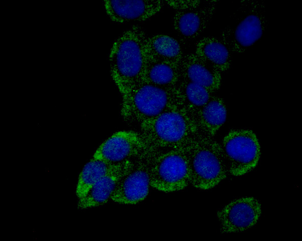
Fig1: ICC staining Emi1 in LOVO cells (green). The nuclear counter stain is DAPI (blue). Cells were fixed in paraformaldehyde, permeabilised with 0.25% Triton X100/PBS.Application
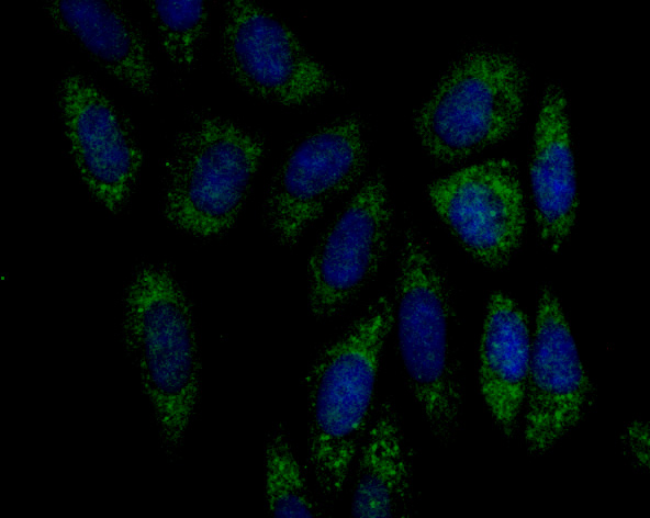
Fig2: ICC staining Emi1 in Siha cells (green). The nuclear counter stain is DAPI (blue). Cells were fixed in paraformaldehyde, permeabilised with 0.25% Triton X100/PBS.Application
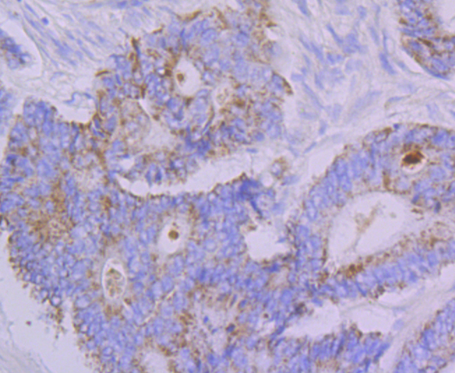
Fig3: Immunohistochemical analysis of paraffin-embedded human colon cancer tissue using anti-Emi1 antibody. Counter stained with hematoxylin.Application
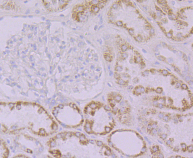
Fig4: Immunohistochemical analysis of paraffin-embedded human kidney tissue using anti-Emi1 antibody. Counter stained with hematoxylin.Application
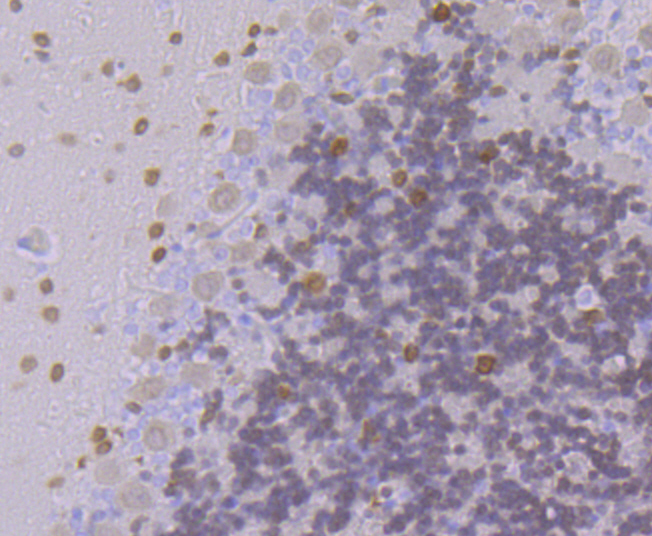
Fig5: Immunohistochemical analysis of paraffin-embedded rat cerebellum tissue using anti-Emi1 antibody. Counter stained with hematoxylin.Application
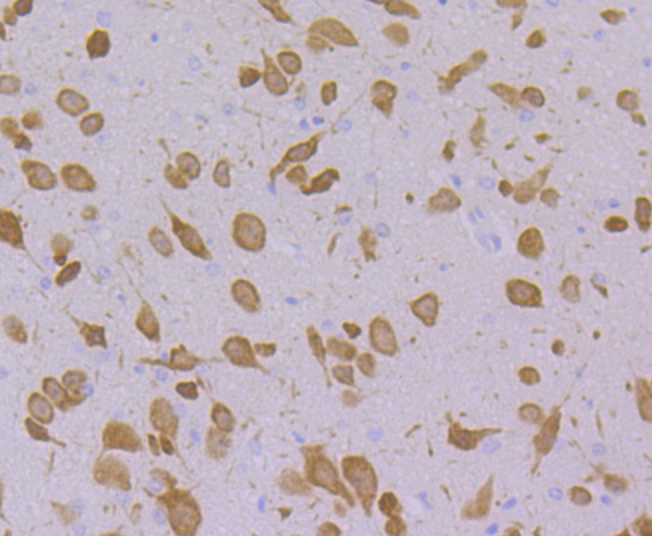
Fig6: Immunohistochemical analysis of paraffin-embedded mouse cerebellum tissue using anti-Emi1 antibody. Counter stained with hematoxylin.Application
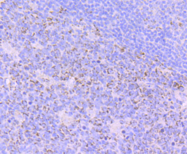
Fig7: Immunohistochemical analysis of paraffin-embedded human tonsil tissue using anti-Emi1 antibody. Counter stained with hematoxylin.| Product Name | Emi1 [JG35-83] |
|---|---|
| Antibody Type | Primary Antibodies |
| Antigen Alias | Early mitotic inhibitor 1 antibody Emi 1 antibody EMI1 antibody F box only protein 5 antibody F box protein 5 antibody F box protein Fbx 5 antibody F box protein Fbx5 antibody F-box only protein 5 antibody FBX5 antibody FBX5_HUMAN antibody Fbxo31 antibody fbxo5 antibody |
| Product description | Emi1 (for early mitotic inhibitor) regulates mitosis by inhibiting the anaphase promoting complex/cyclosome (APC). Emi1 is a conserved F box protein containing a zinc binding region essential for APC inhibition. The Emi1 protein functions to promote cyclin A accumulation and S phase entry in somatic cells by inhibiting the APC complex. At the G1-S transition, Emi1 is transcriptionally induced by the E2F transcription factor. Emi1 overexpression accelerates S phase entry and can override a G1 block caused by overexpression of Cdh1 or the E2F-inhibitor p105 retinoblastoma protein (pRb). Depleting cells of Emi1 through RNA interference prevents accumulation of cyclin A and inhibits S phase entry. Emi1 is required to arrest unfertilized eggs at metaphase of meiosis II and may be the long-sought mediator of CSF activity. Human Emi1 is similar to Xenopus laevis Emi1, which inhibits the APC (Cdc20) ubiquitination complex to allow accumulation of cyclin B. |
| Immunogen | Recombinant protein within human Emi1 aa 50-250. |
| Clonality | Monoclonal |
|---|---|
| Isotype | IgG |
| Host Species | Recombinant rabbit |
| Tested Applications | WBICCIHC |
| ICC:1:50-1:200 IHC:1:50-1:200 WB:1:500 |
|
| Species Reactivity | HumanMouseRat |
| Concentration | 1mg/ml |
| Alternative Names | Early mitotic inhibitor 1 antibody Emi 1 antibody EMI1 antibody F box only protein 5 antibody F box protein 5 antibody F box protein Fbx 5 antibody F box protein Fbx5 antibody F-box only protein 5 antibody FBX5 antibody FBX5_HUMAN antibody Fbxo31 antibody fbxo5 antibody |
|---|---|
| Molecular Weight(MW) | 50 kDa |
| Cellular Localization | Cytoplasm. Cytoskeleton. Nucleus. |
Application
Fig1: ICC staining Emi1 in LOVO cells (green). The nuclear counter stain is DAPI (blue). Cells were fixed in paraformaldehyde, permeabilised with 0.25% Triton X100/PBS.
Application
Fig2: ICC staining Emi1 in Siha cells (green). The nuclear counter stain is DAPI (blue). Cells were fixed in paraformaldehyde, permeabilised with 0.25% Triton X100/PBS.
Application
Fig3: Immunohistochemical analysis of paraffin-embedded human colon cancer tissue using anti-Emi1 antibody. Counter stained with hematoxylin.
Application
Fig4: Immunohistochemical analysis of paraffin-embedded human kidney tissue using anti-Emi1 antibody. Counter stained with hematoxylin.
Application
Fig5: Immunohistochemical analysis of paraffin-embedded rat cerebellum tissue using anti-Emi1 antibody. Counter stained with hematoxylin.
Application
Fig6: Immunohistochemical analysis of paraffin-embedded mouse cerebellum tissue using anti-Emi1 antibody. Counter stained with hematoxylin.
Application
Fig7: Immunohistochemical analysis of paraffin-embedded human tonsil tissue using anti-Emi1 antibody. Counter stained with hematoxylin.| Positive Control | LOVO, Siha, Rat cerebellum tissue, human tonsil tissue, human colon cancer tissue, human kidney tissue, mouse cerebellum tissue. |
|---|---|
| Application Notes | ICC:1:50-1:200 IHC:1:50-1:200 WB:1:500 |
| Form | Liquid |
|---|---|
| Storage Instructions | Store at +4℃ after thawing. Aliquot store at -20℃. Avoid repeated freeze / thaw cycles. |
| Storage Buffer | 1*TBS (pH7.4), 1%BSA, 40%Glycerol. Preservative: 0.05% Sodium Azide. |


