WB
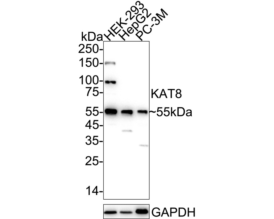
Western blot analysis of KAT8 on different lysates with Rabbit anti-KAT8 antibody at 1/1,000 dilution. Lane 1: HEK-293 cell lysate, Lane 2: HepG2 cell lysate, Lane 3: PC-3M cell lysate, Lysates/proteins at 20 µg/Lane. Exposure time: 3 minutes; 4-20% SDS-PAGE gel. Proteins were transferred to a PVDF membrane and blocked with 5% NFDM/TBST for 1 hour at room temperature. The primary antibody at 1/1,000 dilution was used in 5% NFDM/TBST at 4℃ overnight. Goat Anti-Rabbit IgG - HRP Secondary Antibody at 1/50,000 dilution was used for 1 hour at room temperature.IHC
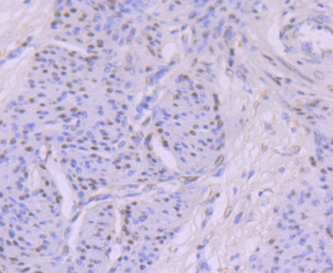
Immunohistochemical analysis of paraffin-embedded human cervix tissue with Rabbit anti-KAT8 antibody at 1/50 dilution. The section was pre-treated using heat mediated antigen retrieval with sodium citrate buffer (pH 6.0) (high pressure) for 2 minutes. The tissues were blocked in 1% BSA for 20 minutes at room temperature, washed with ddH2O and PBS, and then probed with the primary antibody at 1/50 dilution for 1 hour at room temperature. The detection was performed using an HRP conjugated compact polymer system. DAB was used as the chromogen. Tissues were counterstained with hematoxylin and mounted with DPX.IHC
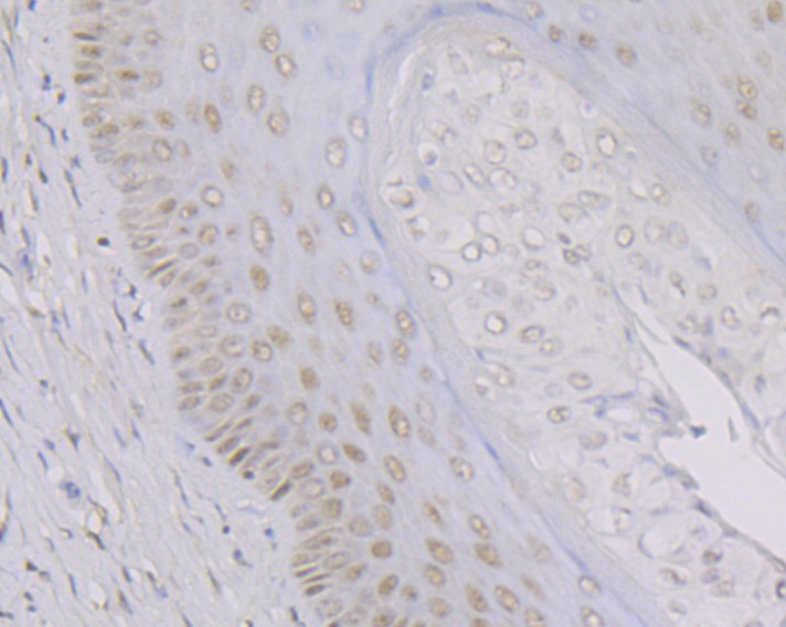
Immunohistochemical analysis of paraffin-embedded rat cervix tissue with Rabbit anti-KAT8 antibody at 1/50 dilution. The section was pre-treated using heat mediated antigen retrieval with sodium citrate buffer (pH 6.0) (high pressure) for 2 minutes. The tissues were blocked in 1% BSA for 20 minutes at room temperature, washed with ddH2O and PBS, and then probed with the primary antibody at 1/50 dilution for 1 hour at room temperature. The detection was performed using an HRP conjugated compact polymer system. DAB was used as the chromogen. Tissues were counterstained with hematoxylin and mounted with DPX.ICC/IF
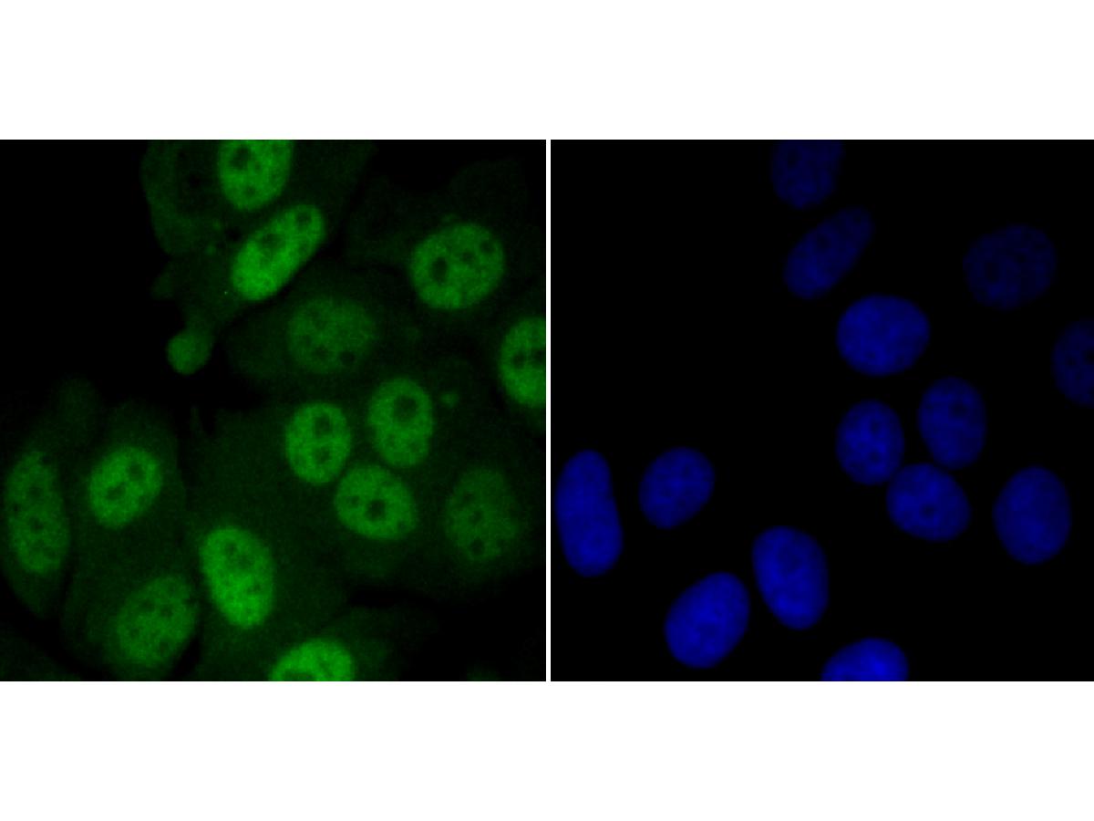
Immunocytochemistry analysis of MCF-7 cells labeling KAT8 with Rabbit anti-KAT8 antibody at 1/50 dilution. Cells were fixed in 4% paraformaldehyde for 10 minutes at 37 ℃, permeabilized with 0.05% Triton X-100 in PBS for 20 minutes, and then blocked with 2% negative goat serum for 30 minutes at room temperature. Cells were then incubated with Rabbit anti-KAT8 antibody at 1/50 dilution in 2% negative goat serum overnight at 4 ℃.Alexa Fluor®488 Goat anti-Rabbit IgG was used as the secondary antibody at 1/1,000 dilution. Nuclear DNA was labelled in blue with DAPI.FC
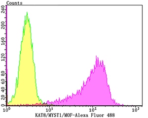
Flow cytometric analysis of K562 cells with KAT8 antibody at 1/100 dilution (yellow) compared with an unlabelled control (cells without incubation with primary antibody; purple).Alexa Fluor 488-conjugated goat anti-rabbit IgG was used as the secondary antibody.| Product Name | KAT8 Recombinant Rabbit Monoclonal Antibody |
|---|---|
| Antibody Type | Primary Antibodies |
| Product description | Dosage compensation ensures that males with a single X chromosome and females with two X chromosomes have the same amount of most X-linked gene products. In Drosophila, this is acheived by enhancing the level of transcription of the X chromosome in males. Proteins such as maleless, male specific lethal 1, 2 and 3, and males absent on the first (MOF) form a dosage compensation complex (DCC) that is required for the twofold increase of transcription of the male X chromosome. The DCC is preferentially associated with many sites on the X chromosome in somatic cells of males. The binding of the DCC to the X chromosome is dependent upon histone 4 acetylation at lysine 16, which is accomplished by MOF. In mammals, MOF (also designated hMOF, MYST1, or MOZ) belongs to the MYST family of histone acetyl transferases which are characterized by a unique C2HC-type zinc finger close to their HAT domains. MOF utilizes the zinc finger domain to contact the globular part of the nucleosome as well as the histone H4 N-terminal tail substrate. The carboxy terminal domain of human MOF also has histone acetyltransferase activity directed against histones H3 and H2A, a characteristic shared with other MYST family histone acetyltransferases. |
| Immunogen | Recombinant protein within Human KAT8 aa 310-458 / 458. |
| Clonality | Monoclonal |
|---|---|
| Isotype | IgG |
| Host Species | Recombinant rabbit |
| Tested Applications | FCICC/IFIHCWB |
| WB:1:1000 IHC:1:50-1:200 ICC/IF:1:50-1:200 FC:1:50-1:100 |
|
| Species Reactivity | HumanMouseRat |
| Concentration | 1mg/ml |
| Purification | Protein A |
| Alternative Names | Histone acetyltransferase KAT8 antibody
Histone acetyltransferase MYST1 antibody
hMOF antibody
K(lysine) acetyltransferase 8 antibody
KAT 8 antibody
Lysine acetyltransferase 8 antibody
MOF antibody
MOZ antibody
MOZ YBF2/SAS3 SAS2 and TIP60 protein 1 antibody MYST 1 antibody MYST histone acetyltransferase 1 antibody myst protein 1 antibody MYST-1 antibody MYST1 antibody MYST1_HUMAN antibody Ortholog of Drosophila males absent on the first (MOF) antibody Probable histone acetyltransferase MYST1 antibody SAS2 and TIP60 protein 1 antibody SAS2 antibody SAS3 antibody TIP60 protein 1 antibody YBF2 antibody YBF2/SAS3 antibody ZC2HC8 antibody |
|---|---|
| Molecular Weight(MW) | 52 kDa(Observed band size: 55 kDa) |
| Cellular Localization | Chromosome. Nucleus. |
| SwissProt ID | Q9H7Z6 |
|---|
WB
Western blot analysis of KAT8 on different lysates with Rabbit anti-KAT8 antibody at 1/1,000 dilution. Lane 1: HEK-293 cell lysate, Lane 2: HepG2 cell lysate, Lane 3: PC-3M cell lysate, Lysates/proteins at 20 µg/Lane. Exposure time: 3 minutes; 4-20% SDS-PAGE gel. Proteins were transferred to a PVDF membrane and blocked with 5% NFDM/TBST for 1 hour at room temperature. The primary antibody at 1/1,000 dilution was used in 5% NFDM/TBST at 4℃ overnight. Goat Anti-Rabbit IgG - HRP Secondary Antibody at 1/50,000 dilution was used for 1 hour at room temperature.
IHC
Immunohistochemical analysis of paraffin-embedded human cervix tissue with Rabbit anti-KAT8 antibody at 1/50 dilution. The section was pre-treated using heat mediated antigen retrieval with sodium citrate buffer (pH 6.0) (high pressure) for 2 minutes. The tissues were blocked in 1% BSA for 20 minutes at room temperature, washed with ddH2O and PBS, and then probed with the primary antibody at 1/50 dilution for 1 hour at room temperature. The detection was performed using an HRP conjugated compact polymer system. DAB was used as the chromogen. Tissues were counterstained with hematoxylin and mounted with DPX.
IHC
Immunohistochemical analysis of paraffin-embedded rat cervix tissue with Rabbit anti-KAT8 antibody at 1/50 dilution. The section was pre-treated using heat mediated antigen retrieval with sodium citrate buffer (pH 6.0) (high pressure) for 2 minutes. The tissues were blocked in 1% BSA for 20 minutes at room temperature, washed with ddH2O and PBS, and then probed with the primary antibody at 1/50 dilution for 1 hour at room temperature. The detection was performed using an HRP conjugated compact polymer system. DAB was used as the chromogen. Tissues were counterstained with hematoxylin and mounted with DPX.
ICC/IF
Immunocytochemistry analysis of MCF-7 cells labeling KAT8 with Rabbit anti-KAT8 antibody at 1/50 dilution. Cells were fixed in 4% paraformaldehyde for 10 minutes at 37 ℃, permeabilized with 0.05% Triton X-100 in PBS for 20 minutes, and then blocked with 2% negative goat serum for 30 minutes at room temperature. Cells were then incubated with Rabbit anti-KAT8 antibody at 1/50 dilution in 2% negative goat serum overnight at 4 ℃.Alexa Fluor®488 Goat anti-Rabbit IgG was used as the secondary antibody at 1/1,000 dilution. Nuclear DNA was labelled in blue with DAPI.
FC
Flow cytometric analysis of K562 cells with KAT8 antibody at 1/100 dilution (yellow) compared with an unlabelled control (cells without incubation with primary antibody; purple).Alexa Fluor 488-conjugated goat anti-rabbit IgG was used as the secondary antibody.| Application Notes | WB:1:1000 IHC:1:50-1:200 ICC/IF:1:50-1:200 FC:1:50-1:100 |
|---|
| Form | Liquid |
|---|---|
| Storage Instructions | Store at +4℃ after thawing. Aliquot store at -20℃. Avoid repeated freeze / thaw cycles. |
| Storage Buffer | 1*TBS (pH7.4), 1%BSA, 40%Glycerol. Preservative: 0.05% Sodium Azide. |


