WB
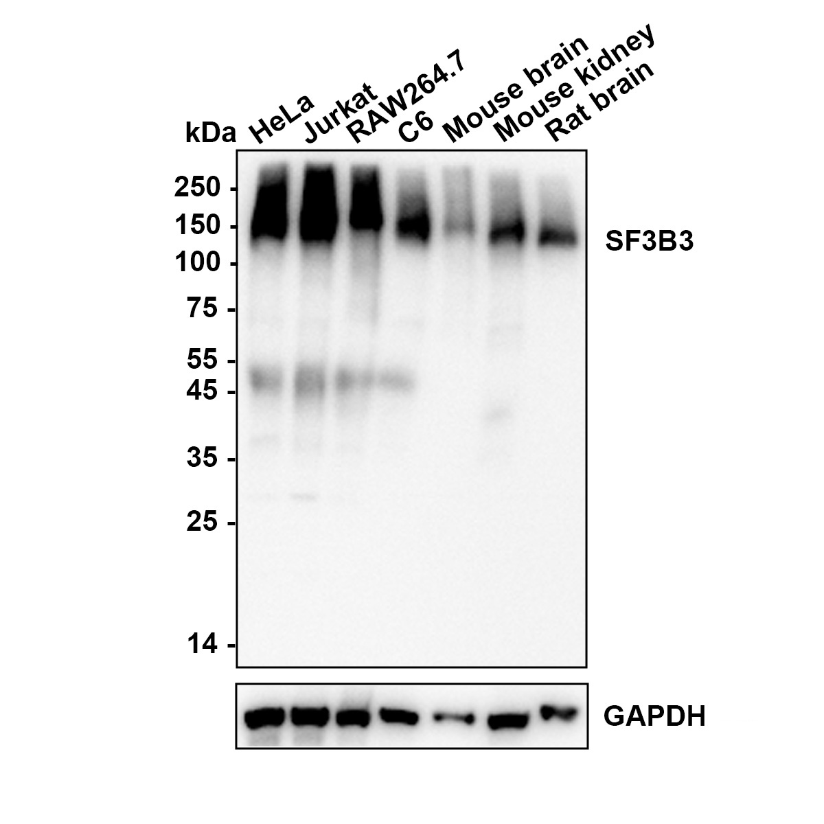
Western blot analysis of SF3B3 on different lysates with Rabbit anti-SF3B3 antibody at 1/5,000 dilution. Lane 1: HeLa, Lane 2: Jurkat, Lane 3: RAW264.7, Lane 4: C6, Lane 5: Mouse brain, Lane 6: Mouse kidney, Lane 7: Rat brain, Lysates/proteins at 20 µg/Lane. Exposure time: 20 seconds; Blocking: 5% NFDM/TBST, 1 hour at room temperature Primary antibody: 1/5,000 in primary antibody dilution buffer, overnight at 4 ℃ Secondary antibody: Goat anti-Rabbit IgG-HRP, 1/50,000 in 5% NFDM/TBST, 1 hour at room temperature.IHC
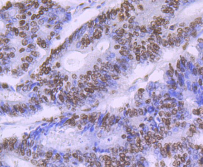
Immunohistochemical analysis of paraffin-embedded human colon cancer tissue using anti-SF3B3 antibody. Counter stained with hematoxylin.IHC
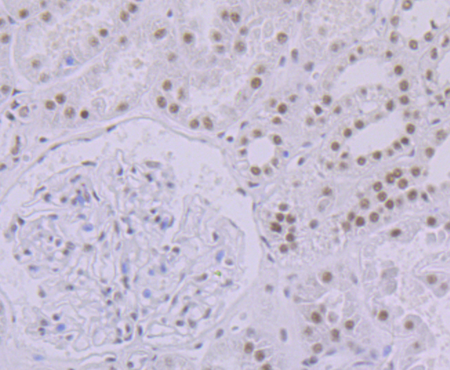
Immunohistochemical analysis of paraffin-embedded human kidney tissue using anti-SF3B3 antibody. Counter stained with hematoxylin.IHC
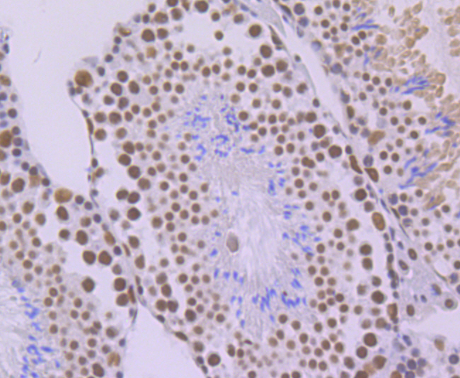
Immunohistochemical analysis of paraffin-embedded mouse testis tissue using anti-SF3B3 antibody. Counter stained with hematoxylin.IHC
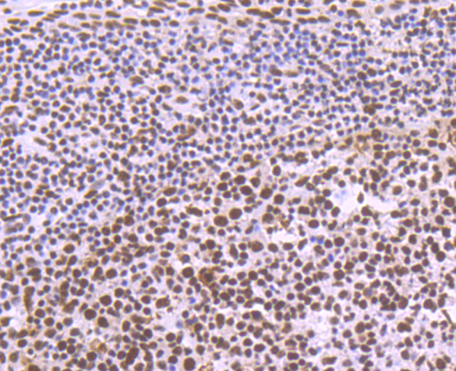
Immunohistochemical analysis of paraffin-embedded human tonsil tissue using anti-SF3B3 antibody. Counter stained with hematoxylin.IHC
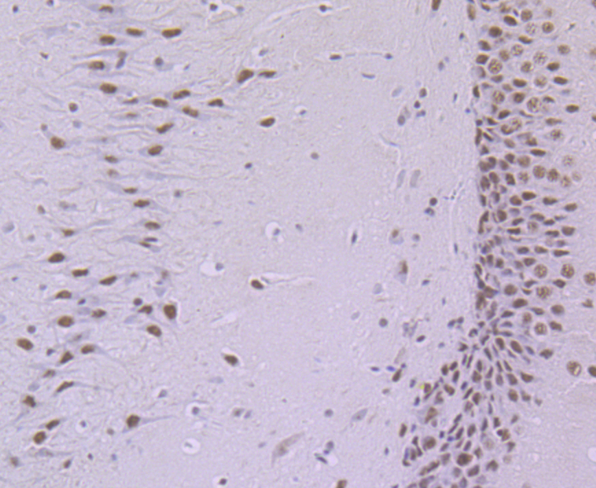
Immunohistochemical analysis of paraffin-embedded rat brain tissue using anti-SF3B3 antibody. Counter stained with hematoxylin.| Product Name | SF3B3 Recombinant Rabbit Monoclonal Antibody |
|---|---|
| Antibody Type | Primary Antibodies |
| Product description | SF3B is a U2 snRNP-associated protein complex essential for spliceosome assembly. SF3B contains the spliceosomal proteins SAP 49, SAP 130 (also known as SF3B3), SAP 145 and SAP 155. SF3B3, SAP 145 and SAP 155 are present in a protein complex in HeLa nuclear extracts and associate with one another. While SF3B3 and SAP 155 interact with each other (directly or indirectly) within this complex, SAP 49 and SAP 145 are known to interact directly with each other. Unexpectedly, the SAP 49-SAP 145 protein-protein interaction requires the amino-terminus of SAP 49, which contains two RNA-recognition motifs. The observation that SAP 49 and SAP 145 interact directly with both U2 snRNP and the pre-mRNA suggests that this protein complex plays a role in tethering U2 snRNP to the branch site. |
| Immunogen | Recombinant protein within Human SF3B3 aa 1061-1217 / 1217. |
| Clonality | Monoclonal |
|---|---|
| Isotype | IgG |
| Host Species | Recombinant rabbit |
| Tested Applications | IHCWB |
| WB:1:500-1:1000 IHC:1:50-1:200 |
|
| Species Reactivity | HumanMouseRat |
| Concentration | 1mg/ml |
| Purification | Protein A |
| Alternative Names | KIAA0017 antibody Pre mRNA splicing factor SF3b 130 kDa subunit antibody Pre-mRNA-splicing factor SF3b 130 kDa subunit antibody RSE1 antibody SAP 130 antibody SAP130 antibody SF3b130 antibody SF3B3 antibody SF3B3_HUMAN antibody Spliceosome associated protein 130 antibody Spliceosome-associated protein 130 antibody Splicing factor 3b subunit 3 130kD antibody Splicing factor 3B subunit 3 antibody STAF130 antibody |
|---|---|
| Molecular Weight(MW) | 136 kDa |
| Cellular Localization | Nucleus. Spliceosome. |
WB
Western blot analysis of SF3B3 on different lysates with Rabbit anti-SF3B3 antibody at 1/5,000 dilution. Lane 1: HeLa, Lane 2: Jurkat, Lane 3: RAW264.7, Lane 4: C6, Lane 5: Mouse brain, Lane 6: Mouse kidney, Lane 7: Rat brain, Lysates/proteins at 20 µg/Lane. Exposure time: 20 seconds; Blocking: 5% NFDM/TBST, 1 hour at room temperature Primary antibody: 1/5,000 in primary antibody dilution buffer, overnight at 4 ℃ Secondary antibody: Goat anti-Rabbit IgG-HRP, 1/50,000 in 5% NFDM/TBST, 1 hour at room temperature.
IHC
Immunohistochemical analysis of paraffin-embedded human colon cancer tissue using anti-SF3B3 antibody. Counter stained with hematoxylin.
IHC
Immunohistochemical analysis of paraffin-embedded human kidney tissue using anti-SF3B3 antibody. Counter stained with hematoxylin.
IHC
Immunohistochemical analysis of paraffin-embedded mouse testis tissue using anti-SF3B3 antibody. Counter stained with hematoxylin.
IHC
Immunohistochemical analysis of paraffin-embedded human tonsil tissue using anti-SF3B3 antibody. Counter stained with hematoxylin.
IHC
Immunohistochemical analysis of paraffin-embedded rat brain tissue using anti-SF3B3 antibody. Counter stained with hematoxylin.| Application Notes | WB:1:500-1:1000 IHC:1:50-1:200 |
|---|
| Form | Liquid |
|---|---|
| Storage Instructions | Store at +4℃ after thawing. Aliquot store at -20℃. Avoid repeated freeze / thaw cycles. |
| Storage Buffer | 1*TBS (pH7.4), 1%BSA, 40%Glycerol. Preservative: 0.05% Sodium Azide. |


