Application
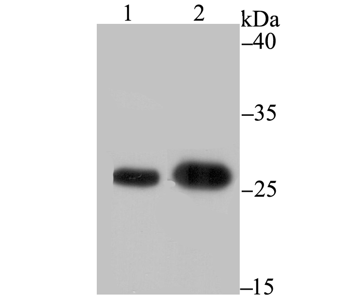
Fig1: Western blot analysis of BAP31 on different lysates using anti-BAP31 antibody at 1/1,000 dilution. Positive control: Lane 1: A431 Lane 2: SK-Br-3Application
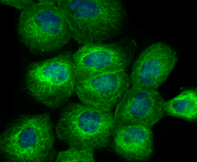
Fig2: ICC staining BAP31 in A431 cells (green). The nuclear counter stain is DAPI (blue). Cells were fixed in paraformaldehyde, permeabilised with 0.25% Triton X100/PBS.Application
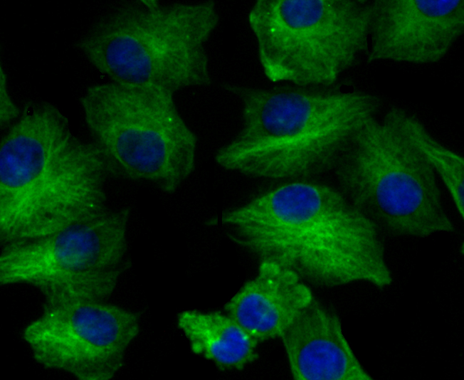
Fig3: ICC staining BAP31 in A549 cells (green). The nuclear counter stain is DAPI (blue). Cells were fixed in paraformaldehyde, permeabilised with 0.25% Triton X100/PBS.Application
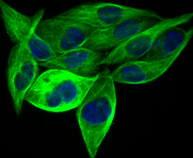
Fig4: ICC staining BAP31 in PC-3M cells (green). The nuclear counter stain is DAPI (blue). Cells were fixed in paraformaldehyde, permeabilised with 0.25% Triton X100/PBS.Application
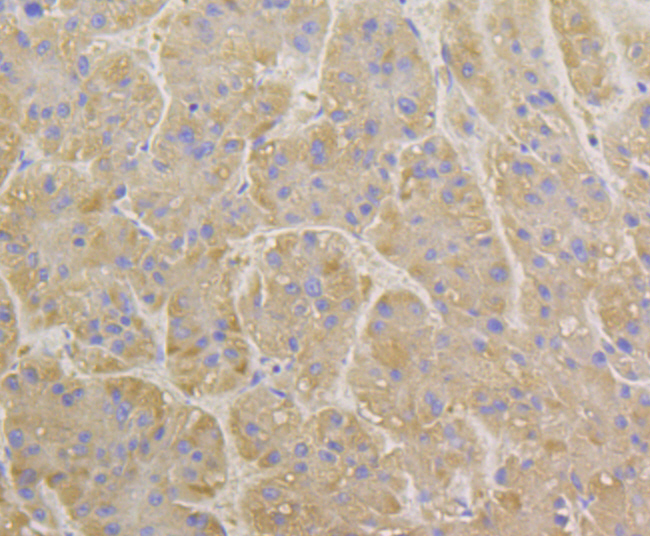
Fig5: Immunohistochemical analysis of paraffin-embedded human liver tissue using anti-BAP31 antibody. Counter stained with hematoxylin.Application
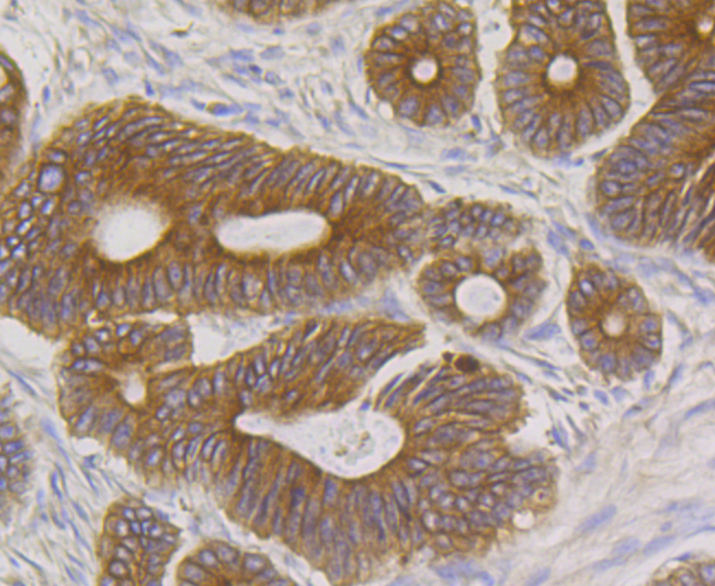
Fig6: Immunohistochemical analysis of paraffin-embedded human colon cancer tissue using anti-BAP31 antibody. Counter stained with hematoxylin.Application
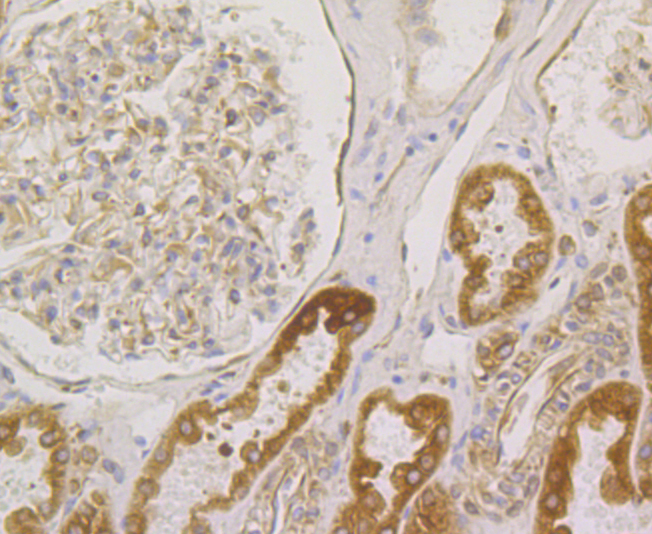
Fig7: Immunohistochemical analysis of paraffin-embedded human kidney tissue using anti-BAP31 antibody. Counter stained with hematoxylin.Application
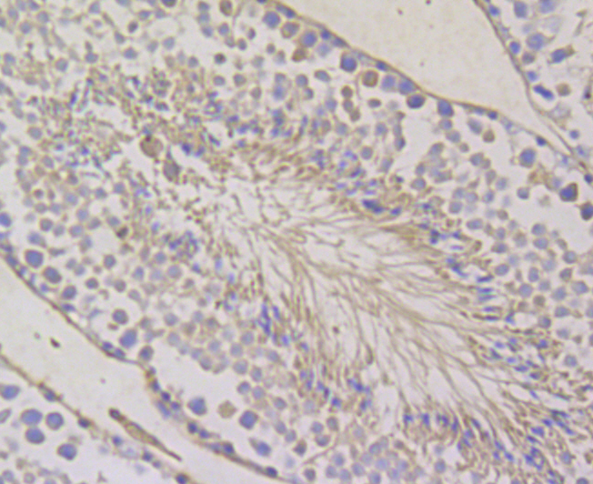
Fig8: Immunohistochemical analysis of paraffin-embedded mouse testis tissue using anti-BAP31 antibody. Counter stained with hematoxylin.Application
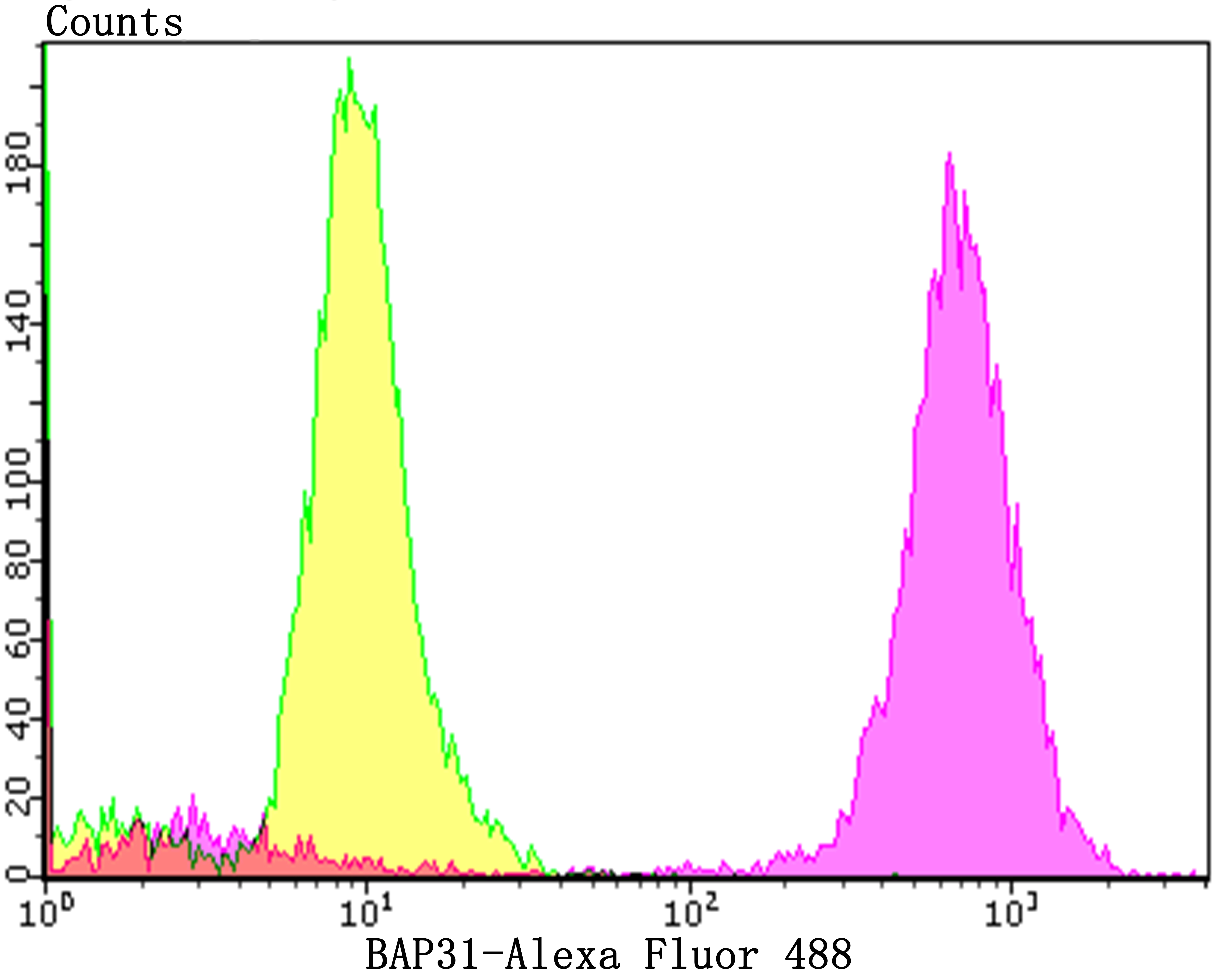
Fig9: Flow cytometric analysis of PC-3M cells with BAP31 antibody at 1/100 dilution (purple) compared with an unlabelled control (cells without incubation with primary antibody; yellow). Alexa Fluor 488-conjugated goat anti-rabbit IgG was used as the secondary antibody.| Product Name | BAP31 [JG37-81] |
|---|---|
| Antibody Type | Primary Antibodies |
| Antigen Alias | 6C6 AG antibody 6C6 AG tumor associated antigen antibody 6C6-AG tumor-associated antigen antibody 6C6AG antibody 6C6AG tumor associated antigen antibody Accessory protein BAP 31 antibody Accessory protein BAP31 antibody B cell receptor associated protein 31 antibody B-cell receptor-associated protein 31 antibody BA31 antibody BAP 31 antibody Bap31 antibody BAP31_HUMAN antibody BCAP 31 antibody BCAP31 antibody BCR associated protein Bap 31 antibody BCR associated protein Bap31 antibody BCR-associated protein 31 antibody CDM antibody CDM protein antibody DXS1357E antibody MS950 antibody p28 antibody p28 Bap31 antibody Protein CDM antibody RP23-329M9.5 antibody |
| Product description | BAP31, a human Bcl-2-interacting protein, is an integral membrane protein that is a component of a protein complex in the endoplasmic reticulum. This protein complex mechanically bridges an apoptosis-initiating caspase, like procaspase-8, with the anti-apoptotic regulator Bcl-2 or Bcl-XL. The cytosolic domain of BAP31 contains two identical caspase recognition sites, which are preferentially cleaved by initiator caspases, including caspase 8. Cleavage of BAP31 during apoptosis generates a p20 fragment, which remains integrated in the membrane and, when expressed ectopically, is a potent inducer of cell death. BAP31 cleavage is important for manifesting cytoplasmic apoptotic events associated with membrane fragmentation and in the cross talk between mitochondria and the endoplasmic reticulum during Fas- mediated apoptosis. The BAP31 gene is ubiquitously expressed in murine tissues and is located on the X chromosome in both mouse and human. |
| Immunogen | Synthetic peptide within the C-terminus of human BAP31. |
| Clonality | Monoclonal |
|---|---|
| Isotype | IgG |
| Host Species | Recombinant rabbit |
| Tested Applications | WBICCIFIHCFC |
| WB:1:500-1:2,000 ICC:1:50-1:200 IHC:1:50-1:200 FC:1:50-1:100 |
|
| Species Reactivity | HumanMouse |
| Concentration | 1mg/ml |
| Alternative Names | 6C6 AG antibody 6C6 AG tumor associated antigen antibody 6C6-AG tumor-associated antigen antibody 6C6AG antibody 6C6AG tumor associated antigen antibody Accessory protein BAP 31 antibody Accessory protein BAP31 antibody B cell receptor associated protein 31 antibody B-cell receptor-associated protein 31 antibody BA31 antibody BAP 31 antibody Bap31 antibody BAP31_HUMAN antibody BCAP 31 antibody BCAP31 antibody BCR associated protein Bap 31 antibody BCR associated protein Bap31 antibody BCR-associated protein 31 antibody CDM antibody CDM protein antibody DXS1357E antibody MS950 antibody p28 antibody p28 Bap31 antibody Protein CDM antibody RP23-329M9.5 antibody |
|---|---|
| Molecular Weight(MW) | 28 kDa |
| Cellular Localization | Endoplasmic reticulum membrane. |
Application
Fig1: Western blot analysis of BAP31 on different lysates using anti-BAP31 antibody at 1/1,000 dilution. Positive control: Lane 1: A431 Lane 2: SK-Br-3
Application
Fig2: ICC staining BAP31 in A431 cells (green). The nuclear counter stain is DAPI (blue). Cells were fixed in paraformaldehyde, permeabilised with 0.25% Triton X100/PBS.
Application
Fig3: ICC staining BAP31 in A549 cells (green). The nuclear counter stain is DAPI (blue). Cells were fixed in paraformaldehyde, permeabilised with 0.25% Triton X100/PBS.
Application
Fig4: ICC staining BAP31 in PC-3M cells (green). The nuclear counter stain is DAPI (blue). Cells were fixed in paraformaldehyde, permeabilised with 0.25% Triton X100/PBS.
Application
Fig5: Immunohistochemical analysis of paraffin-embedded human liver tissue using anti-BAP31 antibody. Counter stained with hematoxylin.
Application
Fig6: Immunohistochemical analysis of paraffin-embedded human colon cancer tissue using anti-BAP31 antibody. Counter stained with hematoxylin.
Application
Fig7: Immunohistochemical analysis of paraffin-embedded human kidney tissue using anti-BAP31 antibody. Counter stained with hematoxylin.
Application
Fig8: Immunohistochemical analysis of paraffin-embedded mouse testis tissue using anti-BAP31 antibody. Counter stained with hematoxylin.
Application
Fig9: Flow cytometric analysis of PC-3M cells with BAP31 antibody at 1/100 dilution (purple) compared with an unlabelled control (cells without incubation with primary antibody; yellow). Alexa Fluor 488-conjugated goat anti-rabbit IgG was used as the secondary antibody.| Positive Control | A431, SK-Br-3, mouse liver tissue lysate, A549, PC-3M, human liver tissue, human colon cancer tissue, human kidney tissue, mouse testis tissue. |
|---|---|
| Application Notes | WB:1:500-1:2,000 ICC:1:50-1:200 IHC:1:50-1:200 FC:1:50-1:100 |
| Form | Liquid |
|---|---|
| Storage Instructions | Store at +4℃ after thawing. Aliquot store at -20℃. Avoid repeated freeze / thaw cycles. |
| Storage Buffer | 1*TBS (pH7.4), 1%BSA, 40%Glycerol. Preservative: 0.05% Sodium Azide. |


