
Application
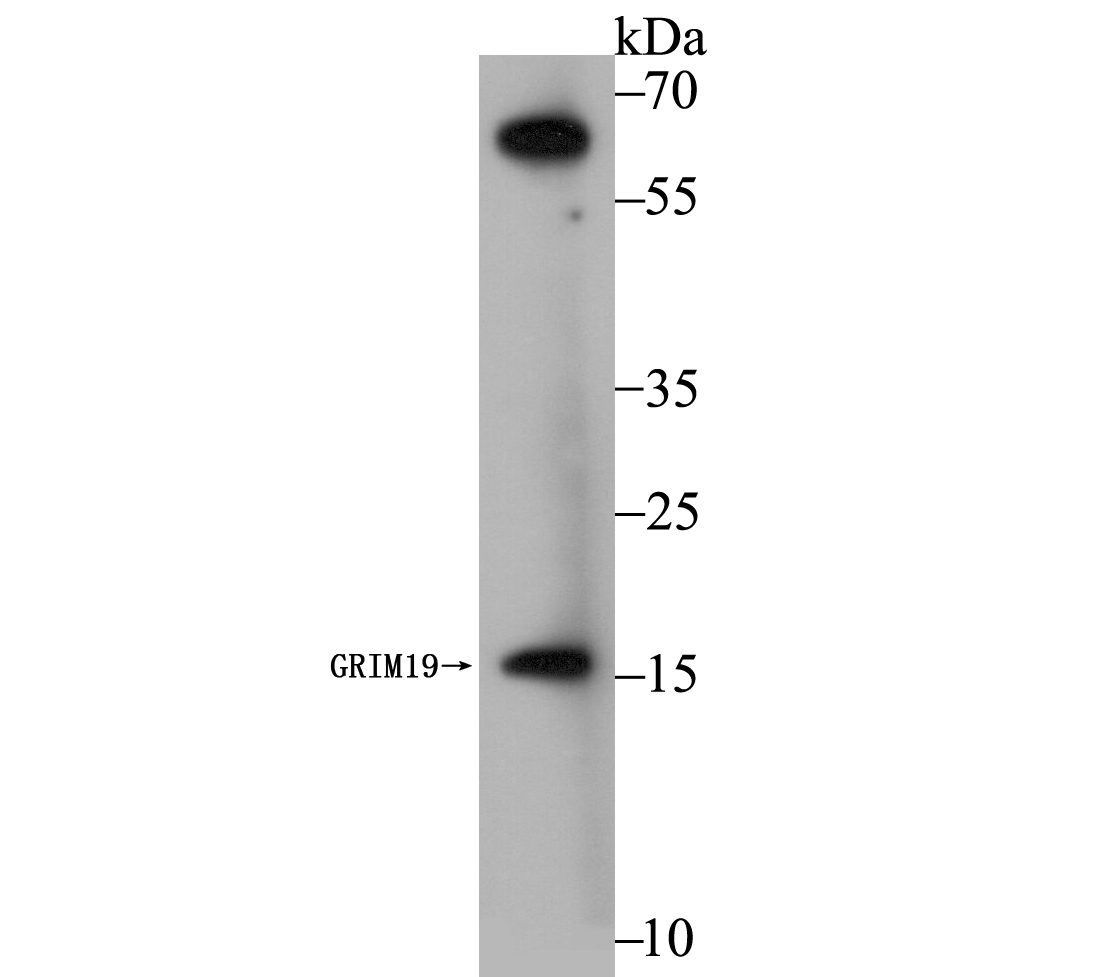
Fig1: Western blot analysis of GRIM19 on MCF-7 cell using anti-GRIM19 antibody at 1/1,000 dilution.Application
Fig2: ICC staining GRIM19 in HepG2 cells (green). The nuclear counter stain is DAPI (blue). Cells were fixed in paraformaldehyde, permeabilised with 0.25% Triton X100/PBS.Application
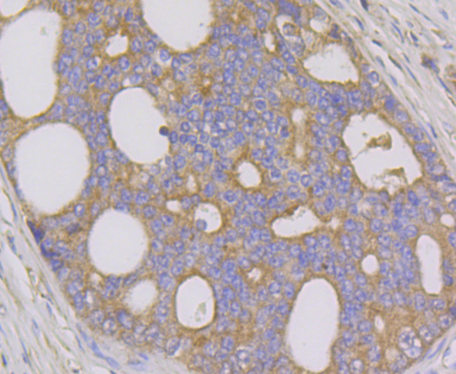
Fig3: Immunohistochemical analysis of paraffin-embedded human prostate cancer tissue using anti-GRIM19 antibody. Counter stained with hematoxylin.Application
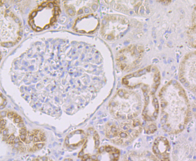
Fig4: Immunohistochemical analysis of paraffin-embedded human kidney tissue using anti-GRIM19 antibody. Counter stained with hematoxylin.Application
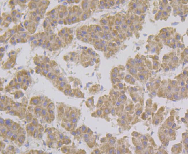
Fig5: Immunohistochemical analysis of paraffin-embedded human liver tissue using anti-GRIM19 antibody. Counter stained with hematoxylin.| Product Name | GRIM19 [JG82-33] |
|---|---|
| Antibody Type | Primary Antibodies |
| Antigen Alias | 2700054G14Rik antibody AU022060 antibody B16.6 antibody CDA016 antibody Cell death regulatory protein antibody Cell death regulatory protein GRIM-19 antibody CGI-39 antibody CGI39 protein antibody CI-B16.6 antibody Complex I-B16.6 antibody FLJ58045 antibody FLJ59191 antibody Gene associated with retinoic and IFN-induced mortality 19 protein antibody Gene associated with retinoic and interferon-induced mortality 19 protein antibody Gene associated with retinoic interferon induced mortality 19 protein antibody GRIM-19 antibody NADH dehydrogenase (ubiquinone) 1 alpha subcomplex, 13 antibody NADH dehydrogenase [ubiquinone] 1 alpha subcomplex subunit 13 antibody NADH ubiquinone oxidoreductase B16.6 subunit antibody NADH-ubiquinone oxidoreductase B16.6 subunit antibody NDUAD_HUMAN antibody NDUFA13 antibody RGD1565358 antibody |
| Product description | The GRIM family of proteins appear to be novel types of tumor suppressors. Grim19, which stands for gene associated with retinoic-interferon-induced mortality 19 protein, is also designated cell death-regulatory protein Grim-19 or NADH dehydrogenase ubiquinone 1 alpha subcomplex subunit 13. The Grim19 protein plays two roles within the cell. As a member of the interferon-beta and retinoic acid-induced pathway of cell death, Grim19 induces apoptosis. As part of the mitochondrial complex I, Grim19 is essential for its assembly and electron transfer activity. It transfers electrons to the respiratory chain from NADH and plays a role in the interferon/all-trans-retinoic acid (IFN/RA) cell death pathway. It localizes primarily to the mitochondrion, but may translocate to the nucleus upon IFN/RA treatment. Grim19 may also be useful as a biological marker or target for drug development. |
| Immunogen | Recombinant protein within human GRIM19 aa 1-150. |
| Clonality | Monoclonal |
|---|---|
| Isotype | IgG |
| Host Species | Recombinant rabbit |
| Tested Applications | WBIHCICC |
| WB:1:500-1:1,000 IHC:1:50-1:200 ICC:1:50-1:200 |
|
| Species Reactivity | Human |
| Concentration | 1mg/ml |
| Alternative Names | 2700054G14Rik antibody
AU022060 antibody
B16.6 antibody
CDA016 antibody
Cell death regulatory protein antibody
Cell death regulatory protein GRIM-19 antibody
CGI-39 antibody
CGI39 protein antibody
CI-B16.6 antibody
Complex I-B16.6 antibody
FLJ58045 antibody
FLJ59191 antibody
Gene associated with retinoic and IFN-induced mortality 19 protein antibody
Gene associated with retinoic and interferon-induced mortality 19 protein antibody
Gene associated with retinoic interferon induced mortality 19 protein antibody
GRIM-19 antibody
NADH dehydrogenase (ubiquinone) 1 alpha subcomplex 13 antibody NADH dehydrogenase [ubiquinone] 1 alpha subcomplex subunit 13 antibody NADH ubiquinone oxidoreductase B16.6 subunit antibody NADH-ubiquinone oxidoreductase B16.6 subunit antibody NDUAD_HUMAN antibody NDUFA13 antibody RGD1565358 antibody |
|---|---|
| Molecular Weight(MW) | 17 kDa |
| Cellular Localization | Mitochondrion. Nucleus. |
| SwissProt ID | Q9P0J0 |
|---|
Application
Fig1: Western blot analysis of GRIM19 on MCF-7 cell using anti-GRIM19 antibody at 1/1,000 dilution.
Application
Fig2: ICC staining GRIM19 in HepG2 cells (green). The nuclear counter stain is DAPI (blue). Cells were fixed in paraformaldehyde, permeabilised with 0.25% Triton X100/PBS.
Application
Fig3: Immunohistochemical analysis of paraffin-embedded human prostate cancer tissue using anti-GRIM19 antibody. Counter stained with hematoxylin.
Application
Fig4: Immunohistochemical analysis of paraffin-embedded human kidney tissue using anti-GRIM19 antibody. Counter stained with hematoxylin.
Application
Fig5: Immunohistochemical analysis of paraffin-embedded human liver tissue using anti-GRIM19 antibody. Counter stained with hematoxylin.| Positive Control | MCF-7, HepG2, human liver tissue, human prostate cancer tissue, human kidney tissue. |
|---|---|
| Application Notes | WB:1:500-1:1,000 IHC:1:50-1:200 ICC:1:50-1:200 |
| Form | Liquid |
|---|---|
| Storage Instructions | Store at +4℃ after thawing. Aliquot store at -20℃. Avoid repeated freeze / thaw cycles. |
| Storage Buffer | 1*TBS (pH7.4), 1%BSA, 40%Glycerol. Preservative: 0.05% Sodium Azide. |


