WB
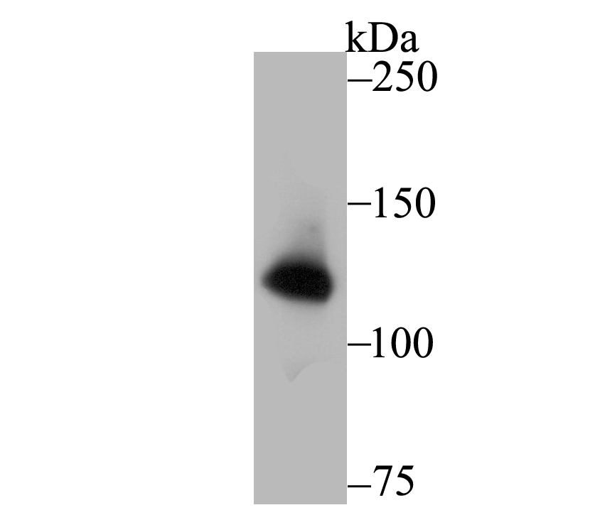
Western blot analysis of hUPF1 on PC-3M cell lysate using anti-hUPF1 antibody at 1/1,000 dilution.ICC/IF
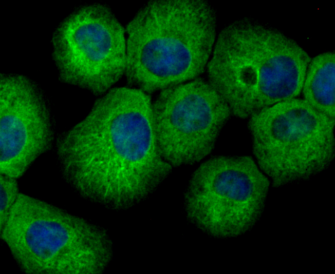
ICC staining hUPF1 in A431 cells (green). The nuclear counter stain is DAPI (blue). Cells were fixed in paraformaldehyde, permeabilised with 0.25% Triton X100/PBS.ICC/IF
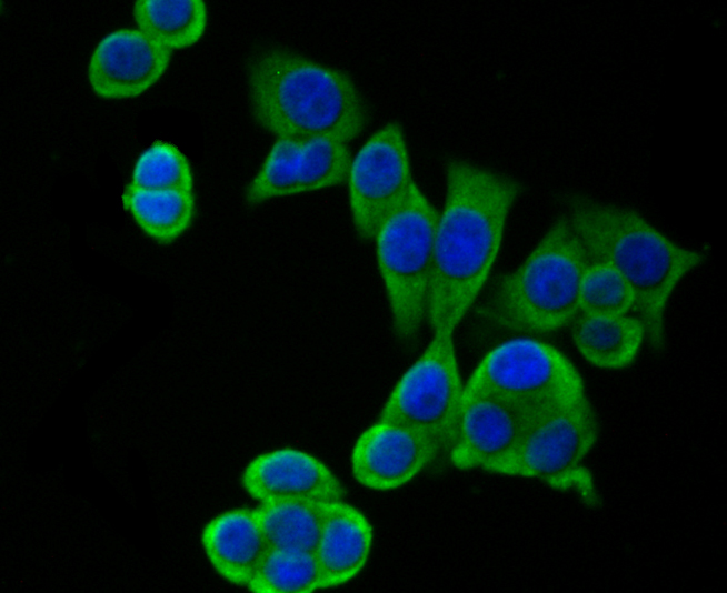
ICC staining hUPF1 in LOVO cells (green). The nuclear counter stain is DAPI (blue). Cells were fixed in paraformaldehyde, permeabilised with 0.25% Triton X100/PBS.ICC/IF
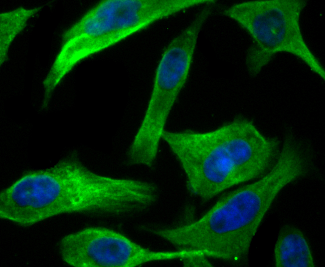
ICC staining hUPF1 in PC-3M cells (green). The nuclear counter stain is DAPI (blue). Cells were fixed in paraformaldehyde, permeabilised with 0.25% Triton X100/PBS.IHC
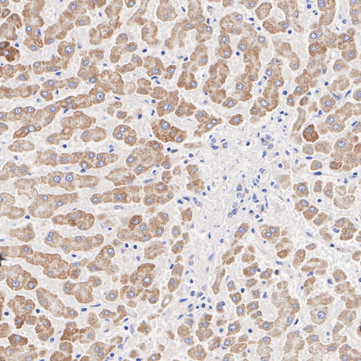
Immunohistochemical analysis of paraffin-embedded human liver tissue with Rabbit anti-hUPF1 antibody at 1/1,000 dilution. The section was pre-treated using heat mediated antigen retrieval with Tris-EDTA buffer (pH 9.0) for 20 minutes. The tissues were blocked in 1% BSA for 20 minutes at room temperature, washed with ddH2O and PBS, and then probed with the primary antibody at 1/1,000 dilution for 1 hour at room temperature. The detection was performed using an HRP conjugated compact polymer system. DAB was used as the chromogen. Tissues were counterstained with hematoxylin and mounted with DPX.IHC
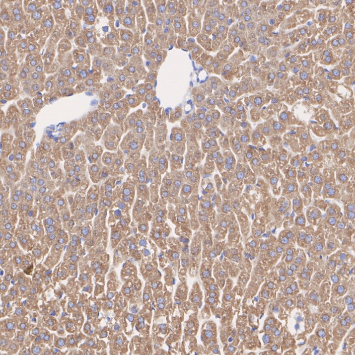
Immunohistochemical analysis of paraffin-embedded mouse liver tissue with Rabbit anti-hUPF1 antibody at 1/1,000 dilution. The section was pre-treated using heat mediated antigen retrieval with Tris-EDTA buffer (pH 9.0) for 20 minutes. The tissues were blocked in 1% BSA for 20 minutes at room temperature, washed with ddH2O and PBS, and then probed with the primary antibody at 1/1,000 dilution for 1 hour at room temperature. The detection was performed using an HRP conjugated compact polymer system. DAB was used as the chromogen. Tissues were counterstained with hematoxylin and mounted with DPX.IHC
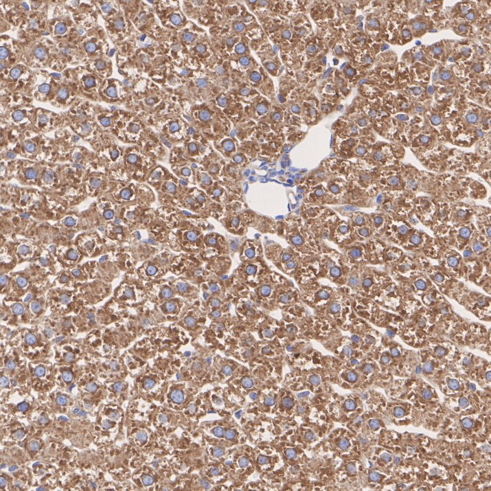
Immunohistochemical analysis of paraffin-embedded rat liver tissue with Rabbit anti-hUPF1 antibody at 1/200 dilution. The section was pre-treated using heat mediated antigen retrieval with Tris-EDTA buffer (pH 9.0) for 20 minutes. The tissues were blocked in 1% BSA for 20 minutes at room temperature, washed with ddH2O and PBS, and then probed with the primary antibody at 1/200 dilution for 1 hour at room temperature. The detection was performed using an HRP conjugated compact polymer system. DAB was used as the chromogen. Tissues were counterstained with hematoxylin and mounted with DPX.FC
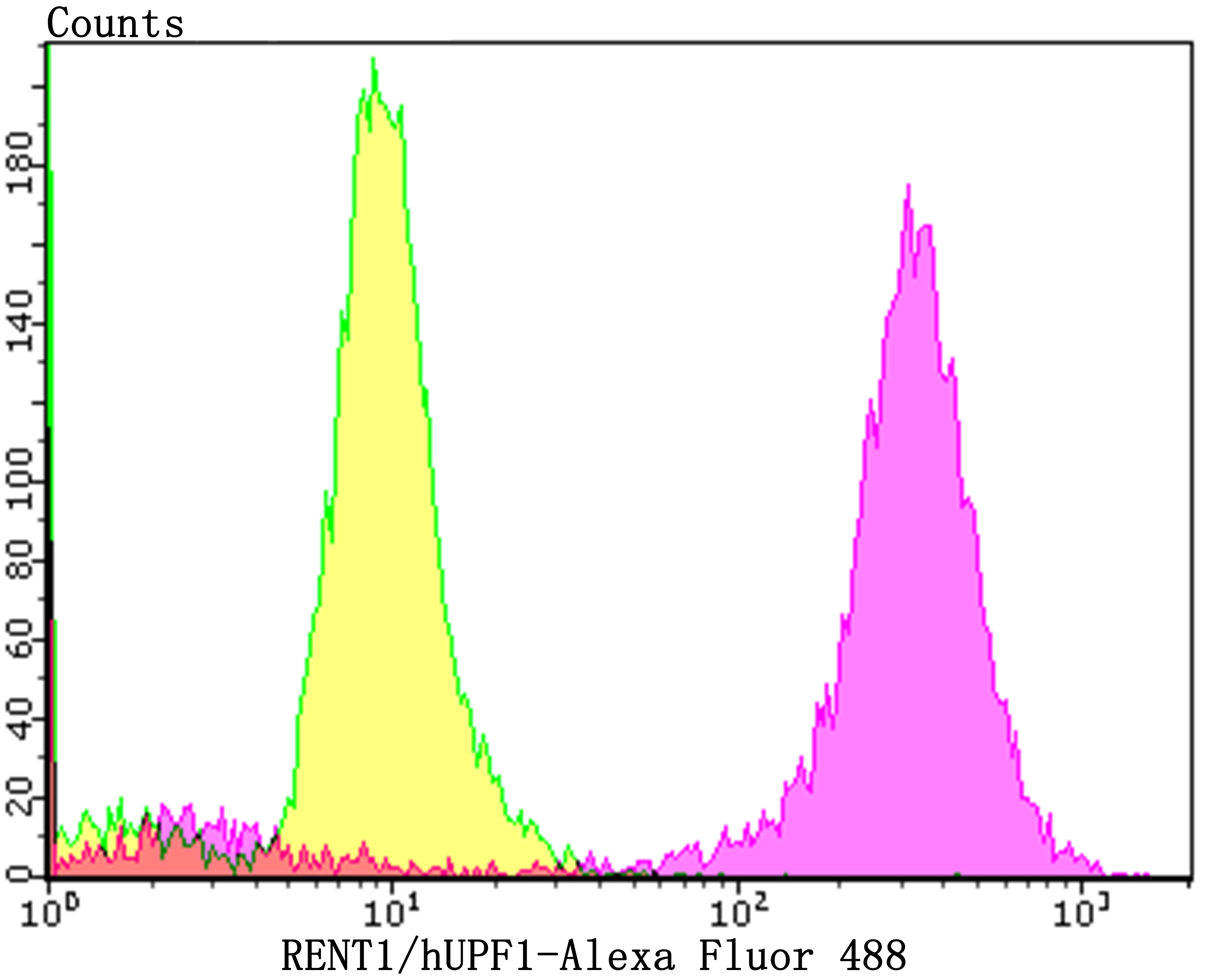
Flow cytometric analysis of PC-3M cells with hUPF1 antibody at 1/100 dilution (purple) compared with an unlabelled control (cells without incubation with primary antibody; yellow). Alexa Fluor 488-conjugated goat anti-rabbit IgG was used as the secondary antibody.| Product Name | hUPF1 Recombinant Rabbit Monoclonal Antibody |
|---|---|
| Antibody Type | Primary Antibodies |
| Product description | In eukaryotes, it is essential to have the ability to detect and degrade transcripts that lack full coding potential. Nonsense-mediated RNA decay (NMD) protects the organism by avoiding the translation of truncated peptides with dominant negative or deleterious gain-of-function potential. Rent1, a mammalian ortholog of Upflp, is essential for embryonic viability. Rent1 (also designated regulator of nonsense transcripts and HUpf1) contains an N-terminal zinc finger-like domain, NTPase domains and a region comprised of domains that define Rent1 as a superfamily group I helicase. Rent1 protein has nucleic-acid-dependent ATPase activity and 5' to 3' helicase activity. In addition, Rent1 is an RNA-binding protein whose activity is modulated by ATP and directly interacts with Rent2, which is a mammalian homolog of Upf2p. Two mammalian orthologs to Upf3p, Rent3a and Rent3b, are encoded by two separate genes. Rent3b (also known as Rent3X) is encoded by a X-linked gene and localizes primarily to the nucleus, while Rent 1 and Rent 2 localize primarily in the cytoplasm. Specific Rent3 protein interactions with Y14 and spliced mRNA suggest Rent3a and Rent3b serve as a link between splicing and NMD in the cytoplasm. |
| Immunogen | Recombinant protein within Human hUPF1 aa 1-120 / 1129. |
| Clonality | Monoclonal |
|---|---|
| Isotype | IgG |
| Host Species | Recombinant rabbit |
| Tested Applications | FCICC/IFIHCWB |
| WB:1:500-1:2000 IHC:1:200-1:1000 ICC/IF:1:50-1:200 FC:1:50-1:100 |
|
| Species Reactivity | HumanMouse |
| Concentration | 1mg/ml |
| Purification | Protein A |
| Alternative Names | ATP dependent helicase RENT1 antibody ATP-dependent helicase RENT1 antibody Delta helicase antibody FLJ43809 antibody FLJ46894 antibody HUPF 1 antibody hUpf1 antibody KIAA0221 antibody Nonsense mRNA reducing factor 1 antibody NORF 1 antibody NORF1 antibody pNORF 1 antibody pNORF1 antibody Regulator of nonsense transcripts 1 antibody RENT 1 antibody RENT1 antibody RENT1_HUMAN antibody Smg 2 antibody Smg 2 homolog nonsense mediated mRNA decay factor antibody UP Frameshift 1 antibody Up frameshift mutation 1 homolog (S. cerevisiae) antibody Up frameshift mutation 1 homolog antibody Up frameshift suppressor 1 homolog antibody Up-frameshift suppressor 1 homolog antibody UPF 1 antibody UPF 1 regulator of nonsense transcripts homolog antibody upf1 antibody UPF1 regulator of nonsense transcripts homolog antibody UPF1 RNA helicase and ATPase antibody Yeast Upf1p homolog antibody |
|---|---|
| Molecular Weight(MW) | 124 kDa |
| Cellular Localization | Cytoplasm. Nucleus. |
WB
Western blot analysis of hUPF1 on PC-3M cell lysate using anti-hUPF1 antibody at 1/1,000 dilution.
ICC/IF
ICC staining hUPF1 in A431 cells (green). The nuclear counter stain is DAPI (blue). Cells were fixed in paraformaldehyde, permeabilised with 0.25% Triton X100/PBS.
ICC/IF
ICC staining hUPF1 in LOVO cells (green). The nuclear counter stain is DAPI (blue). Cells were fixed in paraformaldehyde, permeabilised with 0.25% Triton X100/PBS.
ICC/IF
ICC staining hUPF1 in PC-3M cells (green). The nuclear counter stain is DAPI (blue). Cells were fixed in paraformaldehyde, permeabilised with 0.25% Triton X100/PBS.
IHC
Immunohistochemical analysis of paraffin-embedded human liver tissue with Rabbit anti-hUPF1 antibody at 1/1,000 dilution. The section was pre-treated using heat mediated antigen retrieval with Tris-EDTA buffer (pH 9.0) for 20 minutes. The tissues were blocked in 1% BSA for 20 minutes at room temperature, washed with ddH2O and PBS, and then probed with the primary antibody at 1/1,000 dilution for 1 hour at room temperature. The detection was performed using an HRP conjugated compact polymer system. DAB was used as the chromogen. Tissues were counterstained with hematoxylin and mounted with DPX.
IHC
Immunohistochemical analysis of paraffin-embedded mouse liver tissue with Rabbit anti-hUPF1 antibody at 1/1,000 dilution. The section was pre-treated using heat mediated antigen retrieval with Tris-EDTA buffer (pH 9.0) for 20 minutes. The tissues were blocked in 1% BSA for 20 minutes at room temperature, washed with ddH2O and PBS, and then probed with the primary antibody at 1/1,000 dilution for 1 hour at room temperature. The detection was performed using an HRP conjugated compact polymer system. DAB was used as the chromogen. Tissues were counterstained with hematoxylin and mounted with DPX.
IHC
Immunohistochemical analysis of paraffin-embedded rat liver tissue with Rabbit anti-hUPF1 antibody at 1/200 dilution. The section was pre-treated using heat mediated antigen retrieval with Tris-EDTA buffer (pH 9.0) for 20 minutes. The tissues were blocked in 1% BSA for 20 minutes at room temperature, washed with ddH2O and PBS, and then probed with the primary antibody at 1/200 dilution for 1 hour at room temperature. The detection was performed using an HRP conjugated compact polymer system. DAB was used as the chromogen. Tissues were counterstained with hematoxylin and mounted with DPX.
FC
Flow cytometric analysis of PC-3M cells with hUPF1 antibody at 1/100 dilution (purple) compared with an unlabelled control (cells without incubation with primary antibody; yellow). Alexa Fluor 488-conjugated goat anti-rabbit IgG was used as the secondary antibody.| Application Notes | WB:1:500-1:2000 IHC:1:200-1:1000 ICC/IF:1:50-1:200 FC:1:50-1:100 |
|---|
| Form | Liquid |
|---|---|
| Storage Instructions | Store at +4℃ after thawing. Aliquot store at -20℃. Avoid repeated freeze / thaw cycles. |
| Storage Buffer | 1*TBS (pH7.4), 1%BSA, 40%Glycerol. Preservative: 0.05% Sodium Azide. |


