WB
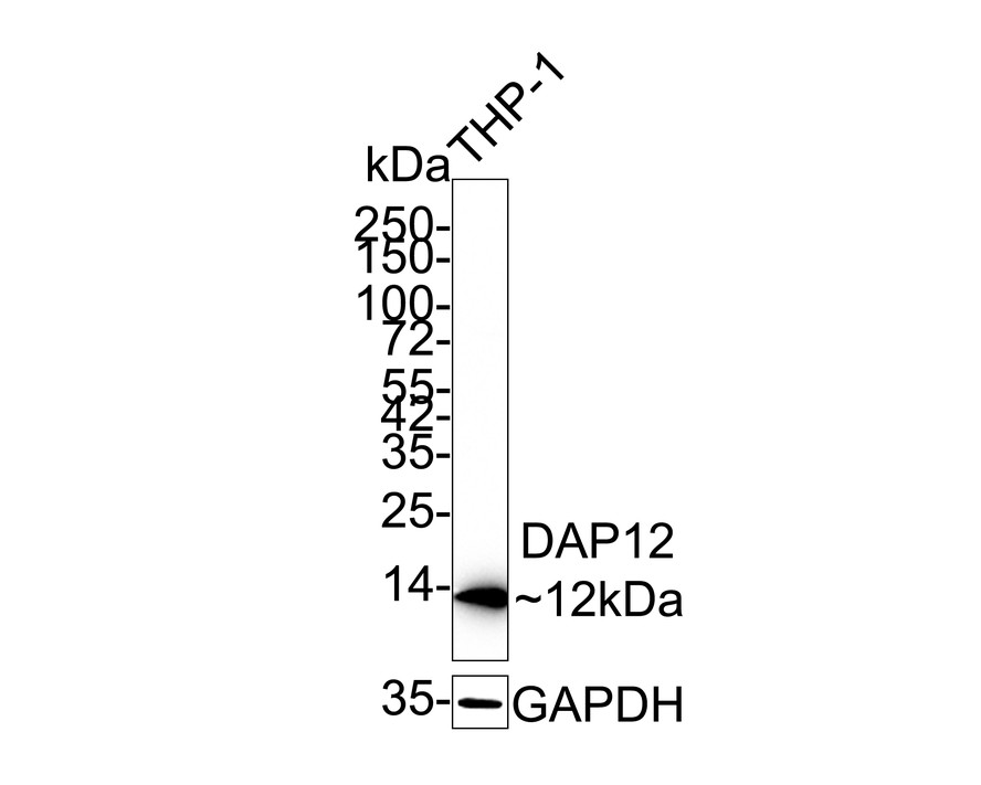
Western blot analysis of DAP12 on THP-1 cell lysates with Rabbit anti-DAP12 antibody at 1/1,000 dilution. Lysates/proteins at 20 µg/Lane. Exposure time: 25 seconds; 4-20% SDS-PAGE gel. Proteins were transferred to a PVDF membrane and blocked with 5% NFDM/TBST for 1 hour at room temperature. The primary antibody at 1/1,000 dilution was used in 5% NFDM/TBST at 4℃ overnight. Goat Anti-Rabbit IgG - HRP Secondary Antibody at 1/50,000 dilution was used for 1 hour at room temperature.IHC
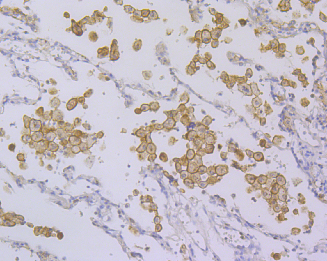
Immunohistochemical analysis of paraffin-embedded human lung cancer tissue using anti-DAP12 antibody. Counter stained with hematoxylin.FC
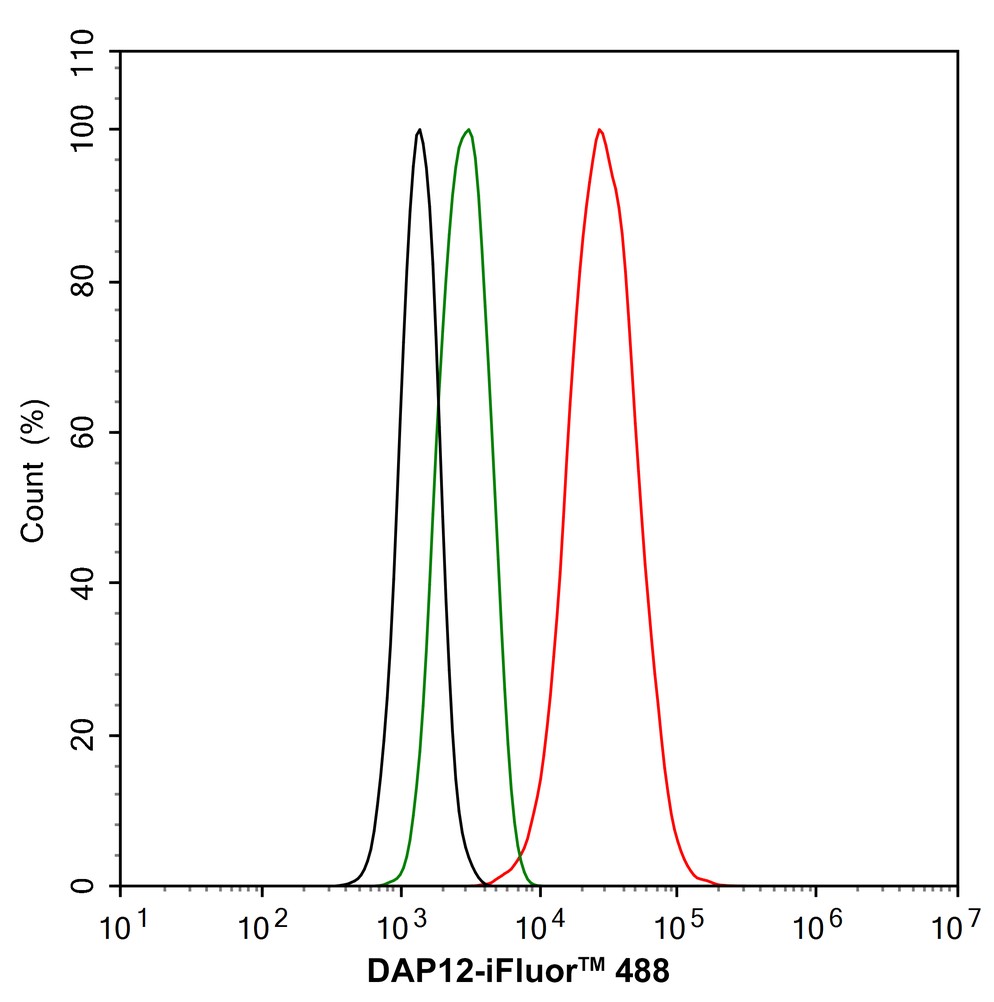
Flow cytometric analysis of THP-1 cells labeling DAP12. Cells were fixed and permeabilized. Then stained with the primary antibody (1μg/mL) (red) compared with Rabbit IgG Isotype Control (green). After incubation of the primary antibody at +4℃ for an hour, the cells were stained with a 488 conjugate-Goat anti-Rabbit IgG Secondary antibody at 1/1,000 dilution for 30 minutes at +4℃. Unlabelled sample was used as a control (cells without incubation with primary antibody; black).| Product Name | DAP12 Recombinant Rabbit Monoclonal Antibody |
|---|---|
| Antibody Type | Primary Antibodies |
| Product description | Natural killer (NK) cells are regulated by stimulatory and inhibitory signals from a variety of receptors. Three main receptor families are responsible for NK cells recognition of MHC I molecules, including Ly-49, CD94/NKG2 and KIR (killer-cell inhibitory receptor). DAP12 is a phosphoprotein that is involved in the activation of NK cells. This protein interacts with membrane glycoproteins of the KIR family, resulting in cellular activation. DAP12 also binds to CD94/NKG2C, an activating NK cell receptor belonging to the C-type lectin superfamily. Additional proteins that bind to DAP12 include Ly-49D and Ly-49H, which associate with DAP12 in the plasma membrane. Phosphorylated DAP12 binds to ZAP-70 and Syk, suggesting that the activation pathway may be similar to that of the T and B cell antigen receptors. |
| Immunogen | Recombinant protein within Human DAP12 aa 1-113 / 113. |
| Clonality | Monoclonal |
|---|---|
| Isotype | IgG |
| Host Species | Recombinant rabbit |
| Tested Applications | FCIHCWB |
| WB:1:500-1:1,000 IHC:1:50-1:200 FC:1:50-1:100 |
|
| Species Reactivity | Human |
| Concentration | 1mg/ml |
| Purification | Protein A |
| Alternative Names | DAP 12 antibody DAP12 antibody DNAX activation protein 12 antibody DNAX-activation protein 12 antibody KAR-associated protein antibody KARAP antibody Killer activating receptor associated protein antibody Killer-activating receptor-associated protein antibody PLOSL antibody TYOBP_HUMAN antibody TYRO protein tyrosine kinase binding protein antibody TYRO protein tyrosine kinase-binding protein antibody TYROBP antibody |
|---|---|
| Molecular Weight(MW) | 12 kDa |
| Cellular Localization | Membrane. |
| SwissProt ID | O43914 |
|---|
WB
Western blot analysis of DAP12 on THP-1 cell lysates with Rabbit anti-DAP12 antibody at 1/1,000 dilution. Lysates/proteins at 20 µg/Lane. Exposure time: 25 seconds; 4-20% SDS-PAGE gel. Proteins were transferred to a PVDF membrane and blocked with 5% NFDM/TBST for 1 hour at room temperature. The primary antibody at 1/1,000 dilution was used in 5% NFDM/TBST at 4℃ overnight. Goat Anti-Rabbit IgG - HRP Secondary Antibody at 1/50,000 dilution was used for 1 hour at room temperature.
IHC
Immunohistochemical analysis of paraffin-embedded human lung cancer tissue using anti-DAP12 antibody. Counter stained with hematoxylin.
FC
Flow cytometric analysis of THP-1 cells labeling DAP12. Cells were fixed and permeabilized. Then stained with the primary antibody (1μg/mL) (red) compared with Rabbit IgG Isotype Control (green). After incubation of the primary antibody at +4℃ for an hour, the cells were stained with a 488 conjugate-Goat anti-Rabbit IgG Secondary antibody at 1/1,000 dilution for 30 minutes at +4℃. Unlabelled sample was used as a control (cells without incubation with primary antibody; black).| Positive Control | THP-1, K562, human lung cancer tissue. |
|---|---|
| Application Notes | WB:1:500-1:1,000 IHC:1:50-1:200 FC:1:50-1:100 |
| Form | Liquid |
|---|---|
| Storage Instructions | Store at +4℃ after thawing. Aliquot store at -20℃. Avoid repeated freeze / thaw cycles. |
| Storage Buffer | 1*TBS (pH7.4), 1%BSA, 40%Glycerol. Preservative: 0.05% Sodium Azide. |


