Application
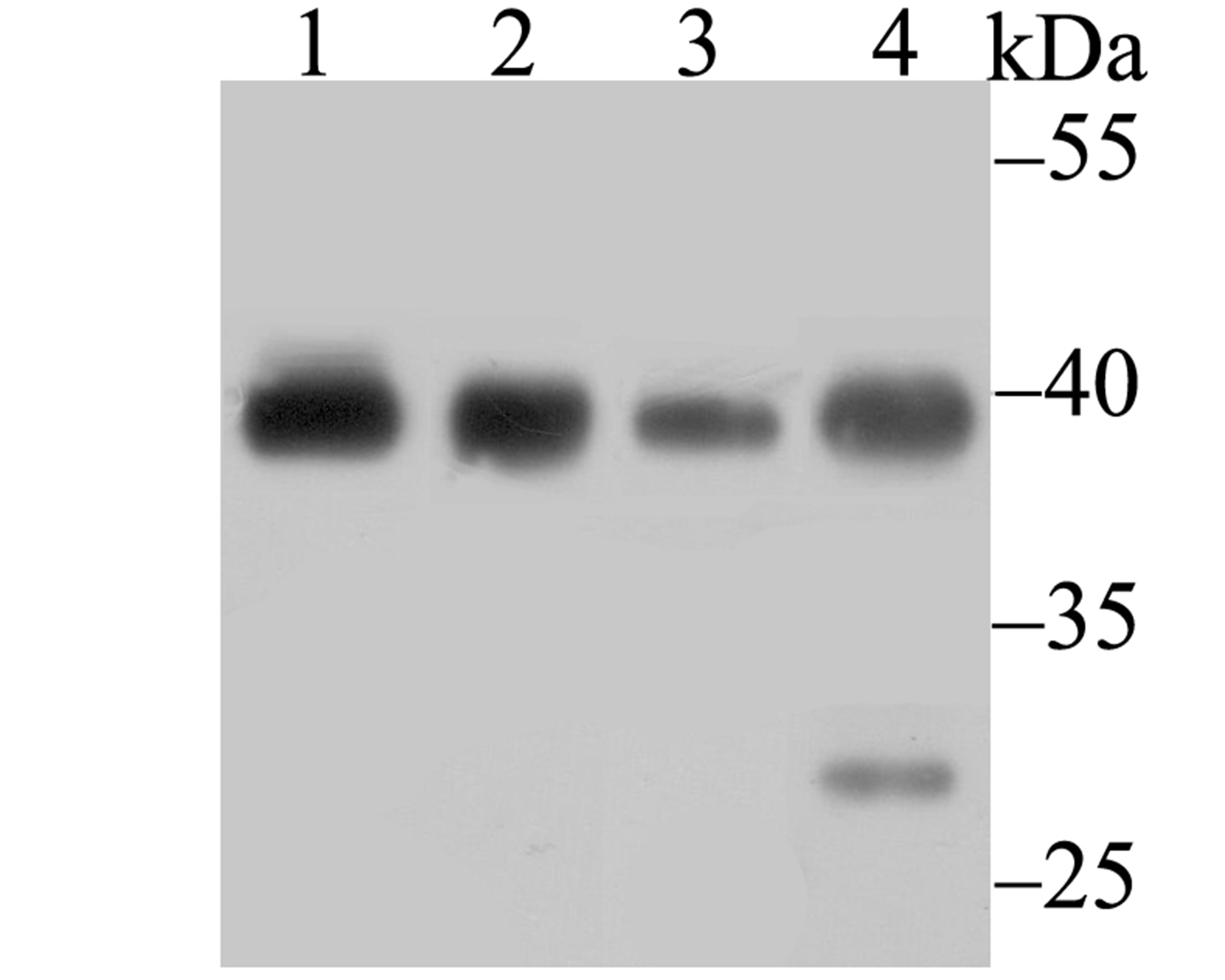
Fig1: Western blot analysis of Bub3 on different lysates using anti-Bub3 antibody at 1/500 dilution. Positive control: Lane 1: A549 Lane 2: HL-60 Lane 3: A431 Lane 4: Rat colonApplication
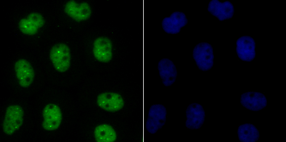
Fig2: ICC staining Bub3 in A431 cells (green). The nuclear counter stain is DAPI (blue). Cells were fixed in paraformaldehyde, permeabilised with 0.25% Triton X100/PBS.Application
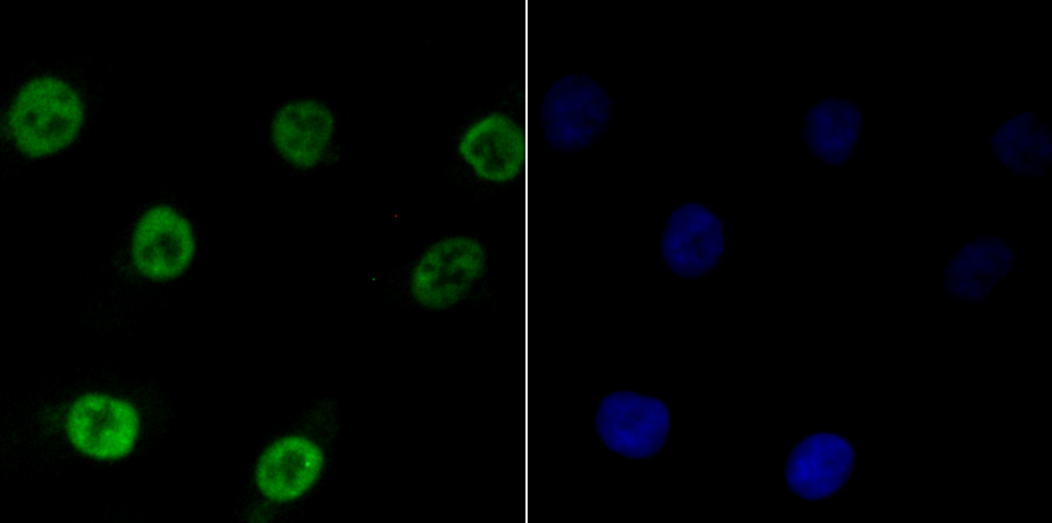
Fig3: ICC staining Bub3 in A549 cells (green). The nuclear counter stain is DAPI (blue). Cells were fixed in paraformaldehyde, permeabilised with 0.25% Triton X100/PBS.Application
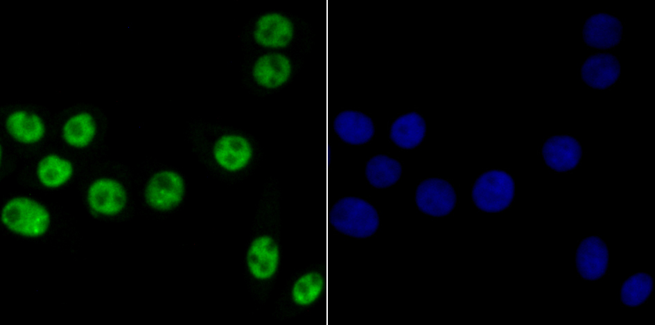
Fig4: ICC staining Bub3 in LOVO cells (green). The nuclear counter stain is DAPI (blue). Cells were fixed in paraformaldehyde, permeabilised with 0.25% Triton X100/PBS.Application
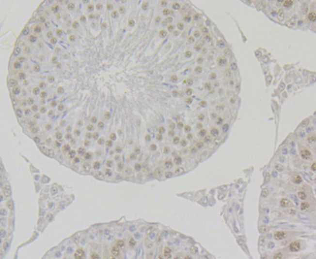
Fig5: Immunohistochemical analysis of paraffin-embedded rat testis tissue using anti-Bub3 antibody. Counter stained with hematoxylin.Application
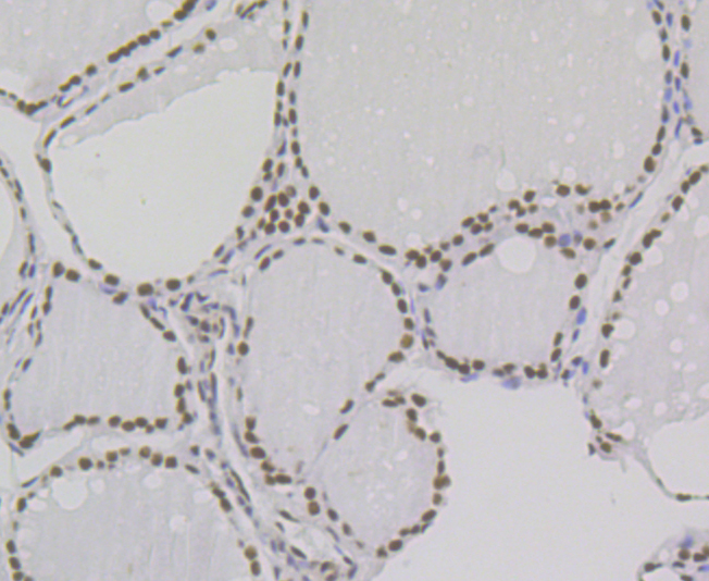
Fig6: Immunohistochemical analysis of paraffin-embedded human thyroid gland tissue using anti-Bub3 antibody. Counter stained with hematoxylin.Application
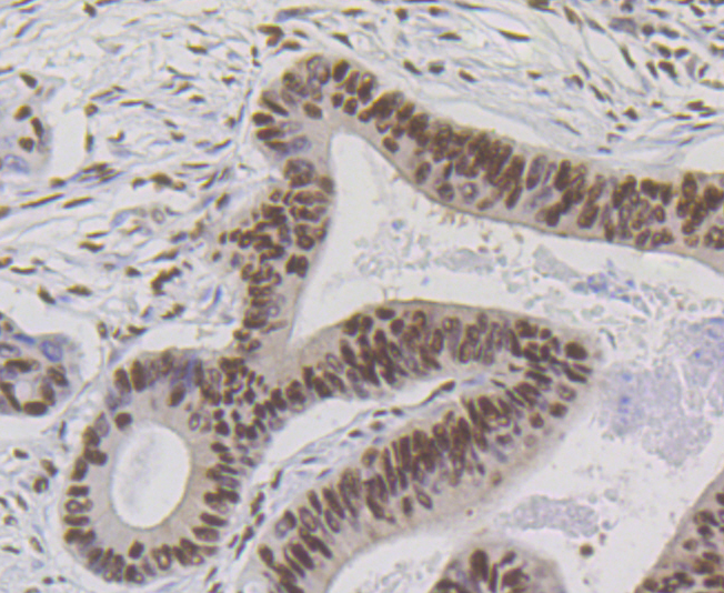
Fig7: Immunohistochemical analysis of paraffin-embedded human colon cancer tissue using anti-Bub3 antibody. Counter stained with hematoxylin.Application
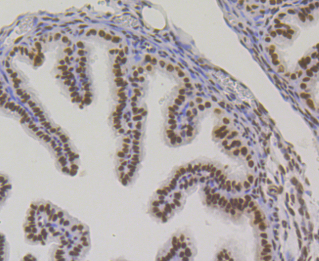
Fig8: Immunohistochemical analysis of paraffin-embedded mouse fallopian tissue using anti-Bub3 antibody. Counter stained with hematoxylin.Application
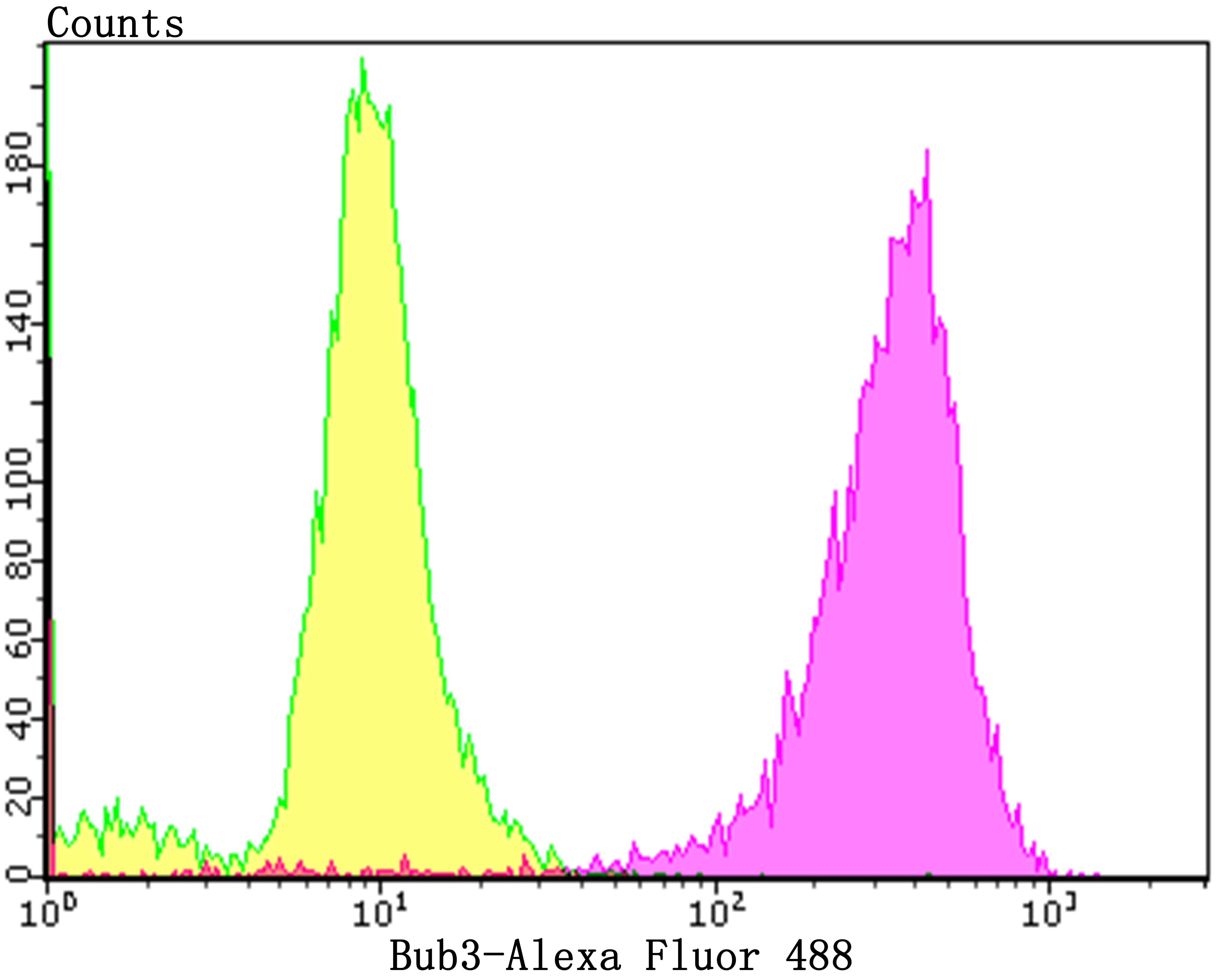
Fig9: Flow cytometric analysis of PC-3M cells with Bub3 antibody at 1/100 dilution (purple) compared with an unlabelled control (cells without incubation with primary antibody; yellow). Alexa Fluor 488-conjugated goat anti-rabbit IgG was used as the secondary antibody.| Product Name | Bub3 [JG38-81] |
|---|---|
| Antibody Type | Primary Antibodies |
| Product description | BUB3 (budding uninhibited by benzimidazoles 3 homolog), also known as BUB3L or hBUB3, is a conserved component of the mitotic spindle assembly complex (MCC). It contains five WD repeat domains and forms cell cycle constitutive complexes with BUB1 and BUBR1. BUB3 is essential for the kinetochore localization of BUB1 and BUBR1. As a component of the MCC, BUB3 is involved in the essential spindle checkpoint pathway that operates during early embryogenesis. The spindle checkpoint pathway functions to postpone the initiation of anaphase until chromosomes are properly attached to the spindle. This acts to ensure accurate chromosome segregation. In addition, BUB3 plays a role in regulating the establishment of correct kinetochore-microtubule attachments. BUB3 is also thought to bind Tctex1L (or DYNLT3), a dynein light chain. |
| Immunogen | Recombinant protein corresponding to the N-terminus of human Bub3. |
| Clonality | Monoclonal |
|---|---|
| Isotype | IgG |
| Host Species | Recombinant rabbit |
| Tested Applications | WB,ICC,IF,IP,IHC,FC |
| WB:1:500-1:2,000 ICC:1:50-1:200 IHC:1:50-1:200 IP:1:10-1:50 FC:1:50-1:100 |
|
| Species Reactivity | Human,Mouse,Rat |
| Concentration | 1mg/ml |
| Purification | Unpurified |
| Alternative Names | Aa2-050 antibody
AU019800 antibody
AU021329 antibody
AU043350 antibody
AW146323 antibody
BUB 3 antibody
BUB 3L antibody
BUB3 (budding uninhibited by benzimidazoles 3 yeast) homolog antibody bub3 antibody BUB3 budding uninhibited by benzimidazoles 3 homolog antibody BUB3 mitotic checkpoint protein antibody BUB3_HUMAN antibody BUB3L antibody Budding uninhibited by benomyl antibody Budding uninhibited by benzimidazoles 3 antibody Budding uninhibited by benzimidazoles 3 homolog (S. cerevisiae) antibody Budding uninhibited by benzimidazoles 3 homolog (yeast) antibody Budding uninhibited by benzimidazoles 3 homolog antibody Budding uninhibited by benzimidazoles 3 S. cerevisiae homolog of antibody C78067 antibody hBUB3 antibody Mitotic checkpoint component antibody Mitotic checkpoint protein bub3 antibody OTTHUMP00000020679 antibody OTTHUMP00000020680 antibody WD repeat type I transmembrane protein A72.5 antibody |
|---|---|
| Molecular Weight(MW) | 37 kDa |
| Cellular Localization | Nucleus. |
Application
Fig1: Western blot analysis of Bub3 on different lysates using anti-Bub3 antibody at 1/500 dilution. Positive control: Lane 1: A549 Lane 2: HL-60 Lane 3: A431 Lane 4: Rat colon
Application
Fig2: ICC staining Bub3 in A431 cells (green). The nuclear counter stain is DAPI (blue). Cells were fixed in paraformaldehyde, permeabilised with 0.25% Triton X100/PBS.
Application
Fig3: ICC staining Bub3 in A549 cells (green). The nuclear counter stain is DAPI (blue). Cells were fixed in paraformaldehyde, permeabilised with 0.25% Triton X100/PBS.
Application
Fig4: ICC staining Bub3 in LOVO cells (green). The nuclear counter stain is DAPI (blue). Cells were fixed in paraformaldehyde, permeabilised with 0.25% Triton X100/PBS.
Application
Fig5: Immunohistochemical analysis of paraffin-embedded rat testis tissue using anti-Bub3 antibody. Counter stained with hematoxylin.
Application
Fig6: Immunohistochemical analysis of paraffin-embedded human thyroid gland tissue using anti-Bub3 antibody. Counter stained with hematoxylin.
Application
Fig7: Immunohistochemical analysis of paraffin-embedded human colon cancer tissue using anti-Bub3 antibody. Counter stained with hematoxylin.
Application
Fig8: Immunohistochemical analysis of paraffin-embedded mouse fallopian tissue using anti-Bub3 antibody. Counter stained with hematoxylin.
Application
Fig9: Flow cytometric analysis of PC-3M cells with Bub3 antibody at 1/100 dilution (purple) compared with an unlabelled control (cells without incubation with primary antibody; yellow). Alexa Fluor 488-conjugated goat anti-rabbit IgG was used as the secondary antibody.| Positive Control | A549, HL-60, A431, rat colon tissue lysate, LOVO, rat testis tissue, human thyroid gland tissue, human colon cancer tissue, mouse fallopian tissue, PC-3M. |
|---|---|
| Application Notes | WB:1:500-1:2,000 ICC:1:50-1:200 IHC:1:50-1:200 IP:1:10-1:50 FC:1:50-1:100 |
| Form | Liquid |
|---|---|
| Storage Instructions | Store at +4℃ after thawing. Aliquot store at -20℃. Avoid repeated freeze / thaw cycles. |
| Storage Buffer | 1*TBS (pH7.4), 1%BSA, 40%Glycerol. Preservative: 0.05% Sodium Azide. |


