WB
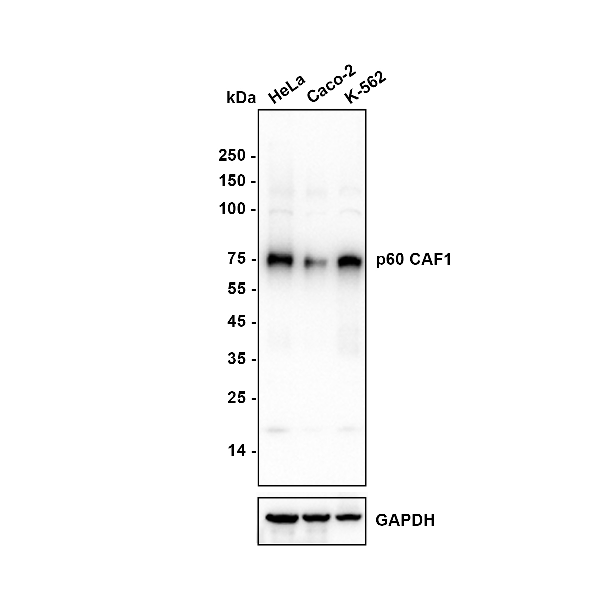
Western blot analysis of p60 CAF1 on different lysates with Rabbit anti-p60 CAF1 antibody at 1/5,000 dilution. Lane 1: HeLa (Human cervical adenocarcinoma cell) cell lysate, Lane 2: Caco-2 (Human colorectal adenocarcinoma cell) cell lysate, Lane 3: K-562 (Human chronic myelogenous leukemia cell) cell lysate, Lysates/proteins at 15 µg/Lane. Exposure time: 10 seconds; Blocking: 5% NFDM/TBST, 1 hour at room temperature Primary antibody: 1/5,000 in primary antibody dilution buffer, overnight at 4 ℃ Secondary antibody: Goat anti-Rabbit IgG-HRP, 1/50,000 in 5% NFDM/TBST, 1 hour at room temperature.IHC
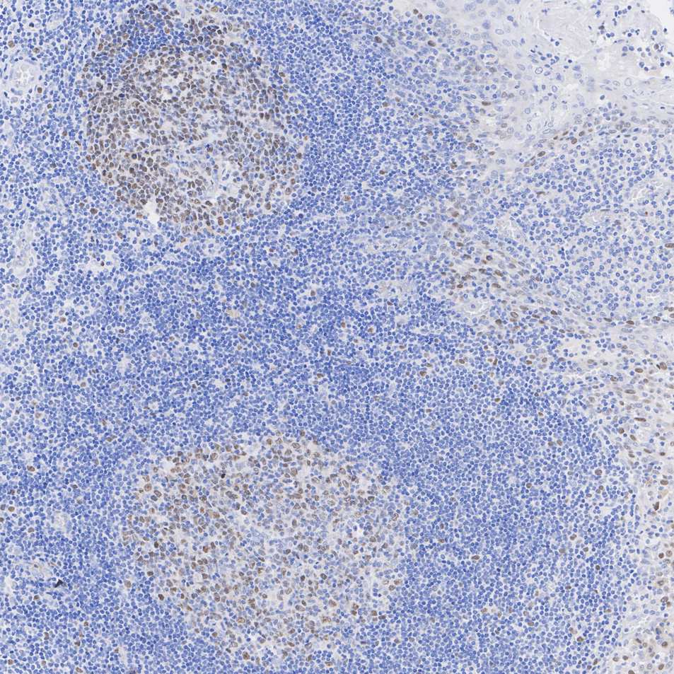
Immunohistochemical analysis of paraffin-embedded human tonsil tissue with Rabbit anti-p60 CAF1 antibody at 1/1,000 dilution. The section was pre-treated using heat mediated antigen retrieval with Tris-EDTA buffer (pH 9.0) for 20 minutes. The tissues were blocked in 1% BSA for 20 minutes at room temperature, washed with ddH2O and PBS, and then probed with the primary antibody at 1/1,000 dilution for 1 hour at room temperature. The detection was performed using an HRP conjugated compact polymer system. DAB was used as the chromogen. Tissues were counterstained with hematoxylin and mounted with DPX.IHC
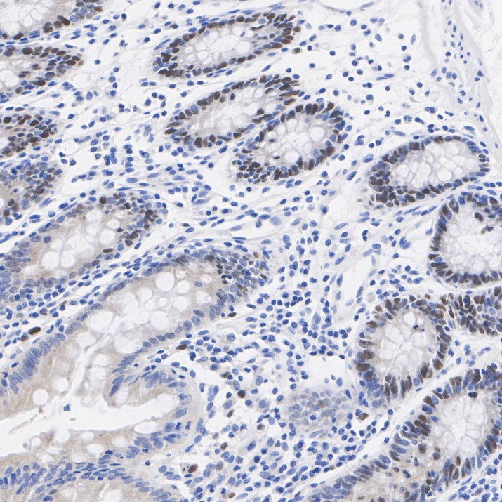
Immunohistochemical analysis of paraffin-embedded human colon tissue with Rabbit anti-p60 CAF1 antibody at 1/1,000 dilution. The section was pre-treated using heat mediated antigen retrieval with Tris-EDTA buffer (pH 9.0) for 20 minutes. The tissues were blocked in 1% BSA for 20 minutes at room temperature, washed with ddH2O and PBS, and then probed with the primary antibody at 1/1,000 dilution for 1 hour at room temperature. The detection was performed using an HRP conjugated compact polymer system. DAB was used as the chromogen. Tissues were counterstained with hematoxylin and mounted with DPX.| Product Name | p60 CAF1 Recombinant Rabbit Monoclonal Antibody |
|---|---|
| Antibody Type | Primary Antibodies |
| Product description | Chromatin assembly factor-1 (CAF-1) is a multisubunit protein complex that comprises three polypeptide subunits known as p150, p60, and p48. CAF-1 is a nucleosome assembly factor that deposits newly synthesized and acetylated histones H3/H4 into nascent chromatin during DNA replication. The p150 subunit of CAF-1 also supports the maintenance of heterochromatin, which requires the synthesis of both new histones and heterochromatin proteins and their orderly assembly during DNA replication. Heterochromatin is characterized as densely coiled chromatin that generally replicates late during S phase, has a low gene density, and contains large blocks of repetitive DNA that is relatively inaccessible to DNA-modifying reagents. In late S phase, p150 directly associates with heterochromatin associated proteins 1 (HP1α, HP1β and HP1γ). As cells prepare for mitosis, CAF-1 p150 and some HP1 progressively dissociate from heterochromatin, coinciding with the phosphorylation of histone H3. The HP1 proteins reassociate with chromatin at the end of mitosis, as histone H3 is dephosphorylated. |
| Immunogen | Recombinant protein within Human p60 CAF1 aa 419-559 / 559. |
| Clonality | Monoclonal |
|---|---|
| Isotype | IgG |
| Host Species | Recombinant rabbit |
| Tested Applications | FCIHCWB |
| WB:1:5000 IHC:1:1000 |
|
| Species Reactivity | Human |
| Concentration | 1mg/ml |
| Purification | Protein A |
| Alternative Names | CAF 1 subunit B antibody CAF 1A antibody CAF I 60 kDa subunit antibody CAF IP60 antibody CAF-1 antibody CAF-1 subunit B antibody CAF-I 60 kDa subunit antibody CAF-I p60 antibody CAF1 antibody CAF1A antibody CAF1B_HUMAN antibody CAF1P60 antibody CHAF 1B antibody CHAF1B antibody Chromatin assembly factor 1 subunit B antibody Chromatin assembly factor I antibody Chromatin assembly factor I p60 subunit antibody Human chromatin assembly factor-I p60 subunit antibody M phase phosphoprotein 7 antibody M-phase phosphoprotein 7 antibody MPHOSPH7 antibody MPP 7 antibody MPP7 antibody p60 subunit antibody |
|---|---|
| Molecular Weight(MW) | 61 kDa(Observed band size: 70 kDa) |
| Cellular Localization | Cytoplasm. Nucleus. |
| SwissProt ID | Q13112 |
|---|
WB
Western blot analysis of p60 CAF1 on different lysates with Rabbit anti-p60 CAF1 antibody at 1/5,000 dilution. Lane 1: HeLa (Human cervical adenocarcinoma cell) cell lysate, Lane 2: Caco-2 (Human colorectal adenocarcinoma cell) cell lysate, Lane 3: K-562 (Human chronic myelogenous leukemia cell) cell lysate, Lysates/proteins at 15 µg/Lane. Exposure time: 10 seconds; Blocking: 5% NFDM/TBST, 1 hour at room temperature Primary antibody: 1/5,000 in primary antibody dilution buffer, overnight at 4 ℃ Secondary antibody: Goat anti-Rabbit IgG-HRP, 1/50,000 in 5% NFDM/TBST, 1 hour at room temperature.
IHC
Immunohistochemical analysis of paraffin-embedded human tonsil tissue with Rabbit anti-p60 CAF1 antibody at 1/1,000 dilution. The section was pre-treated using heat mediated antigen retrieval with Tris-EDTA buffer (pH 9.0) for 20 minutes. The tissues were blocked in 1% BSA for 20 minutes at room temperature, washed with ddH2O and PBS, and then probed with the primary antibody at 1/1,000 dilution for 1 hour at room temperature. The detection was performed using an HRP conjugated compact polymer system. DAB was used as the chromogen. Tissues were counterstained with hematoxylin and mounted with DPX.
IHC
Immunohistochemical analysis of paraffin-embedded human colon tissue with Rabbit anti-p60 CAF1 antibody at 1/1,000 dilution. The section was pre-treated using heat mediated antigen retrieval with Tris-EDTA buffer (pH 9.0) for 20 minutes. The tissues were blocked in 1% BSA for 20 minutes at room temperature, washed with ddH2O and PBS, and then probed with the primary antibody at 1/1,000 dilution for 1 hour at room temperature. The detection was performed using an HRP conjugated compact polymer system. DAB was used as the chromogen. Tissues were counterstained with hematoxylin and mounted with DPX.| Application Notes | WB:1:5000 IHC:1:1000 |
|---|
| Form | Liquid |
|---|---|
| Storage Instructions | Store at +4℃ after thawing. Aliquot store at -20℃. Avoid repeated freeze / thaw cycles. |
| Storage Buffer | 1*TBS (pH7.4), 1%BSA, 40%Glycerol. Preservative: 0.05% Sodium Azide. |


