Application
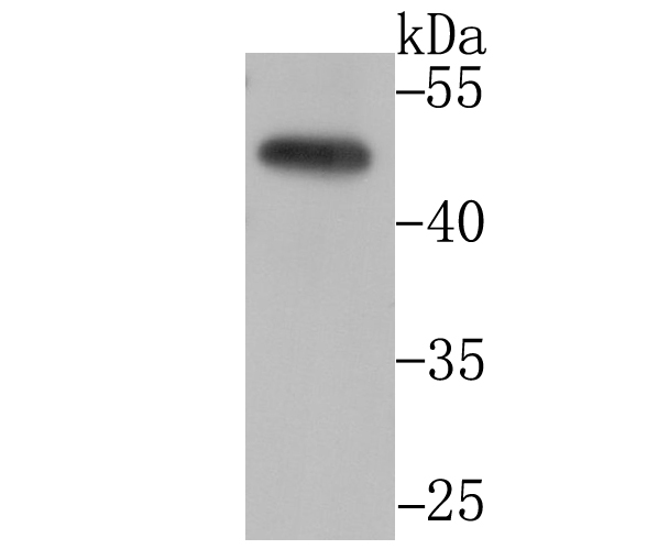
Fig1: Western blot analysis of CtBP1 on PC-3M cell lysate using anti-CtBP1 antibody at 1/1,000 dilution.Application
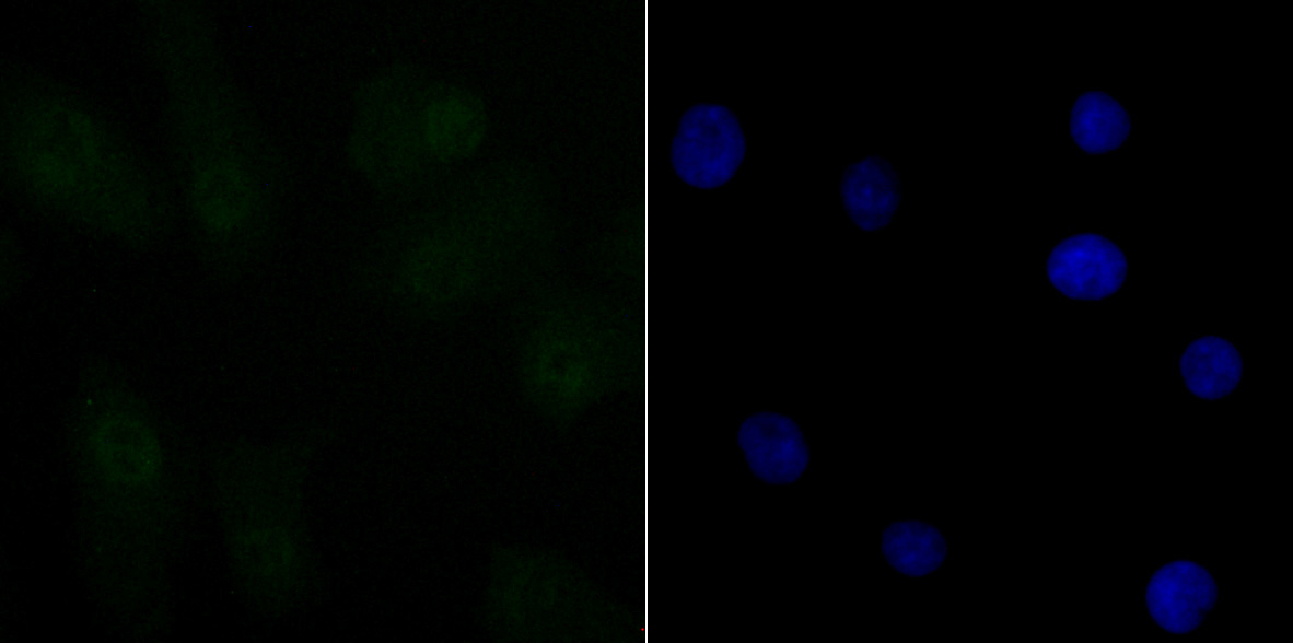
Fig2: ICC staining CtBP1 in A549 cells (green). The nuclear counter stain is DAPI (blue). Cells were fixed in paraformaldehyde, permeabilised with 0.25% Triton X100/PBS.Application
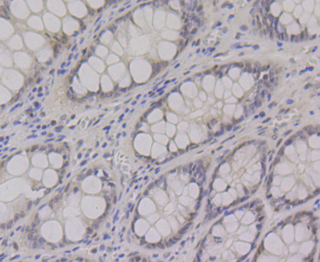
Fig3: Immunohistochemical analysis of paraffin-embedded human colon tissue using anti-CtBP1 antibody. Counter stained with hematoxylin.Application
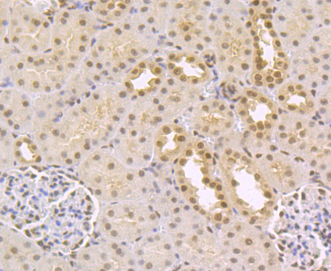
Fig4: Immunohistochemical analysis of paraffin-embedded rat kidney tissue using anti-CtBP1 antibody. Counter stained with hematoxylin.| Product Name | CtBP1 [JG39-73] |
|---|---|
| Antibody Type | Primary Antibodies |
| Antigen Alias | BARS antibody brefeldin A- ribosylated substrate antibody C terminal binding protein 1 antibody C-terminal-binding protein 1 antibody CTBP antibody CtBP1 antibody CTBP1_HUMAN antibody MGC104684 antibody |
| Product description | otein that associates with various proteins and functions as a corepressor of transcription. CtBP1 and the related protein CtBP2 are characterized as C-terminal binding protein of adenovirus E1A, and they preferentially associate with the E1A via a 5-amino acid motif, PLDLS, to repress E1A induced oncogenesis and cellular transformation. CtBP1 is expressed from embryo to adult, but CtBP2 is mainly expressed during embryogenesis. During skeletal and T-cell development, CtBP1 and CtBP2 associate with the PLDLSL domain of δEF1, a cellular zinc finger-homeodomain protein, and thereby enhances δEF1 induced transcriptional silencing. In addition, CtBP complexes with CtIP, a protein that recognizes distinctly different protein motifs from CtBP. CtIP binds to the BRCT repeats within the breast cancer gene BRCA1 and enables CtBP to influence BRCA1 activity. CtIP/CtBP binding to BRCA1 inhibits the transactivation of the p21 promoter, and it is critical for regulating p21 transcription in response to DNA damage. |
| Immunogen | Recombinant protein within human CtBP1 aa 300-500. |
| Clonality | Monoclonal |
|---|---|
| Isotype | IgG |
| Host Species | Recombinant rabbit |
| Tested Applications | WBIHCICCIFIP |
| WB:1:500-1:2,000 IHC:1:50-1:200 ICC:1:50-1:100 IP:1:10-1:50 |
|
| Species Reactivity | HumanMouseRat |
| Concentration | 1mg/ml |
| Alternative Names | BARS antibody brefeldin A- ribosylated substrate antibody C terminal binding protein 1 antibody C-terminal-binding protein 1 antibody CTBP antibody CtBP1 antibody CTBP1_HUMAN antibody MGC104684 antibody |
|---|---|
| Molecular Weight(MW) | 47 kDa |
| Cellular Localization | Cytoplasm. Nucleus. |
Application
Fig1: Western blot analysis of CtBP1 on PC-3M cell lysate using anti-CtBP1 antibody at 1/1,000 dilution.
Application
Fig2: ICC staining CtBP1 in A549 cells (green). The nuclear counter stain is DAPI (blue). Cells were fixed in paraformaldehyde, permeabilised with 0.25% Triton X100/PBS.
Application
Fig3: Immunohistochemical analysis of paraffin-embedded human colon tissue using anti-CtBP1 antibody. Counter stained with hematoxylin.
Application
Fig4: Immunohistochemical analysis of paraffin-embedded rat kidney tissue using anti-CtBP1 antibody. Counter stained with hematoxylin.| Positive Control | PC-3M, rat kidney tissue, human colon tissue, A549. |
|---|---|
| Application Notes | WB:1:500-1:2,000 IHC:1:50-1:200 ICC:1:50-1:100 IP:1:10-1:50 |
| Form | Liquid |
|---|---|
| Storage Instructions | Store at +4℃ after thawing. Aliquot store at -20℃. Avoid repeated freeze / thaw cycles. |
| Storage Buffer | 1*TBS (pH7.4), 1%BSA, 40%Glycerol. Preservative: 0.05% Sodium Azide. |


