WB
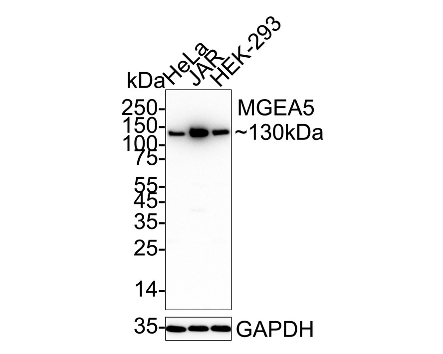
Western blot analysis of MGEA5 on different lysates with Rabbit anti-MGEA5 antibody at 1/1,000 dilution. Lane 1: HeLa cell lysate, Lane 2: JAR cell lysate, Lane 3: HEK-293 cell lysate, Lysates/proteins at 20 µg/Lane. Exposure time: 30 seconds; 4-20% SDS-PAGE gel. Proteins were transferred to a PVDF membrane and blocked with 5% NFDM/TBST for 1 hour at room temperature. The primary antibody at 1/1,000 dilution was used in 5% NFDM/TBST at 4℃ overnight. Goat Anti-Rabbit IgG - HRP Secondary Antibody at 1/50,000 dilution was used for 1 hour at room temperature.IHC
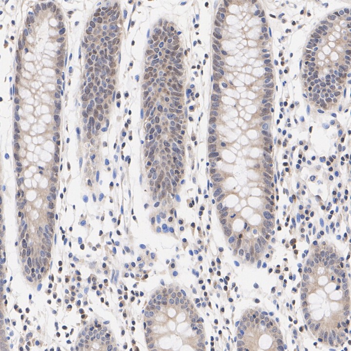
Immunohistochemical analysis of paraffin-embedded human colon tissue with Rabbit anti-MGEA5 antibody at 1/1,000 dilution. The section was pre-treated using heat mediated antigen retrieval with Tris-EDTA buffer (pH 9.0) for 20 minutes. The tissues were blocked in 1% BSA for 20 minutes at room temperature, washed with ddH2O and PBS, and then probed with the primary antibody at 1/1,000 dilution for 1 hour at room temperature. The detection was performed using an HRP conjugated compact polymer system. DAB was used as the chromogen. Tissues were counterstained with hematoxylin and mounted with DPX.IHC
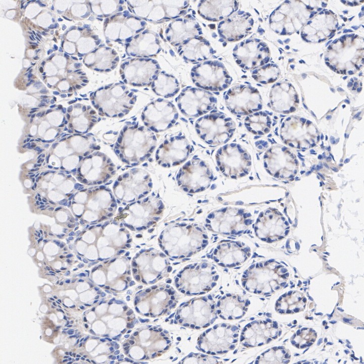
Immunohistochemical analysis of paraffin-embedded mouse colon tissue with Rabbit anti-MGEA5 antibody at 1/200 dilution. The section was pre-treated using heat mediated antigen retrieval with Tris-EDTA buffer (pH 9.0) for 20 minutes. The tissues were blocked in 1% BSA for 20 minutes at room temperature, washed with ddH2O and PBS, and then probed with the primary antibody at 1/200 dilution for 1 hour at room temperature. The detection was performed using an HRP conjugated compact polymer system. DAB was used as the chromogen. Tissues were counterstained with hematoxylin and mounted with DPX.IHC
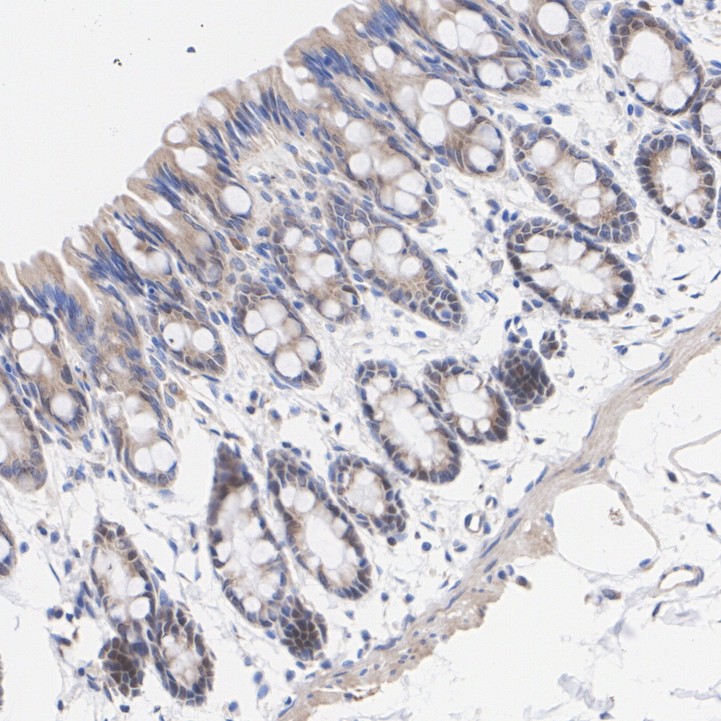
Immunohistochemical analysis of paraffin-embedded rat colon tissue with Rabbit anti-MGEA5 antibody at 1/200 dilution. The section was pre-treated using heat mediated antigen retrieval with Tris-EDTA buffer (pH 9.0) for 20 minutes. The tissues were blocked in 1% BSA for 20 minutes at room temperature, washed with ddH2O and PBS, and then probed with the primary antibody at 1/200 dilution for 1 hour at room temperature. The detection was performed using an HRP conjugated compact polymer system. DAB was used as the chromogen. Tissues were counterstained with hematoxylin and mounted with DPX.IF-P
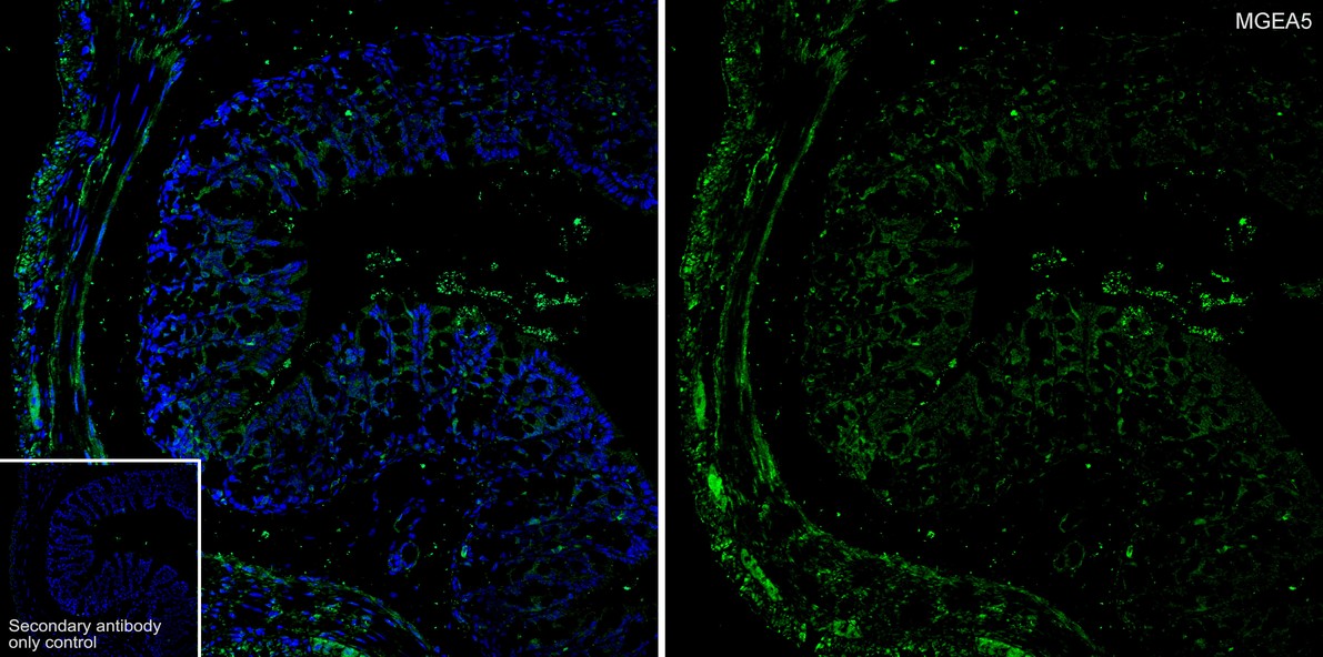
Species: Mouse, Site: colon, Sample: Paraffin-embedded section, Antibody concentration: 1/200.IP
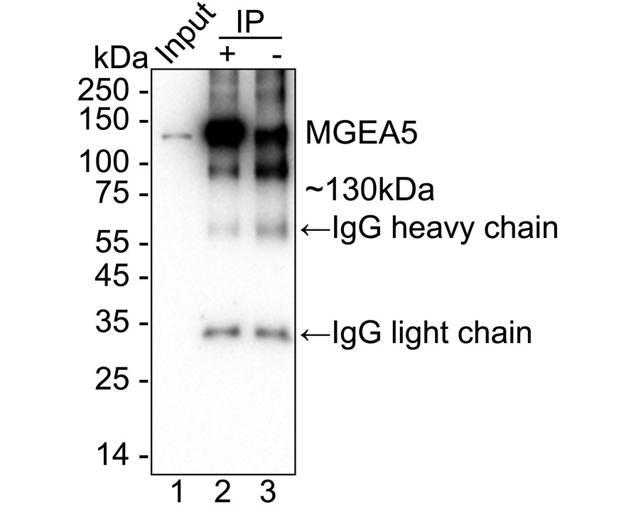
MGEA5 was immunoprecipitated from 0.2 mg HeLa cell lysate with Rabbit anti-MGEA5 antibody at 2 µg/10 µl beads. Western blot was performed from the immunoprecipitate using Rabbit anti-MGEA5 antibody at 1/5,000 dilution. Anti-Rabbit IgG for IP Nano-secondary antibody at 1/5,000 dilution was used for 1 hour at room temperature. Lane 1: HeLa cell lysate (input), Lane 2: Rabbit anti-MGEA5 antibody IP in HeLa cell lysate, Lane 3: Rabbit IgG instead of Rabbit anti-MGEA5 antibody in HeLa cell lysate, Blocking/Dilution buffer: 5% NFDM/TBST, Exposure time: 30 seconds.| Product Name | MGEA5 Recombinant Rabbit Monoclonal Antibody |
|---|---|
| Antibody Type | Primary Antibodies |
| Product description | Cleaves GlcNAc but not GalNAc from O-glycosylated proteins. Can use p-nitrophenyl-beta-GlcNAc and 4-methylumbelliferone-GlcNAc as substrates but not p-nitrophenyl-beta-GalNAc or p-nitrophenyl-alpha-GlcNAc (in vitro). Does not bind acetyl-CoA and does not have histone acetyltransferase activity. Cleaves GlcNAc but not GalNAc from O-glycosylated proteins. Can use p-nitrophenyl-beta-GlcNAc as substrate but not p-nitrophenyl-beta-GalNAc or p-nitrophenyl-alpha-GlcNAc (in vitro), but has about six times lower specific activity than isoform 1. |
| Immunogen | Synthetic peptide within Human MGEA5 aa 2-103 / 916. |
| Clonality | Monoclonal |
|---|---|
| Isotype | IgG |
| Host Species | Recombinant rabbit |
| Tested Applications | IF-PIHCIPWB |
| WB:1:1000-1:2000 IHC:1:50-1:200 IF-P:1:200 IP:1-2μg/sample |
|
| Species Reactivity | HumanMouseRat |
| Concentration | 1mg/ml |
| Purification | Protein A |
| Alternative Names | Beta-hexosaminidase antibody Beta-N-acetylglucosaminidase antibody Beta-N-acetylhexosaminidase antibody Bifunctional protein NCOAT antibody HEXC antibody Hyaluronidase in meningioma antibody KIAA0679 antibody MEA5 antibody Meningioma expressed antigen 5 antibody Meningioma-expressed antigen 5 antibody MGEA 5 antibody Mgea5 antibody N-acetyl-beta-D-glucosaminidase antibody N-acetyl-beta-glucosaminidase antibody NCOAT antibody Nuclear cytoplasmic O GlcNAcase and acetyltransferase antibody Nuclear cytoplasmic O-GlcNAcase and acetyltransferase antibody OGA antibody OGA_HUMAN antibody Protein O-GlcNAcase antibody |
|---|---|
| Molecular Weight(MW) | 103 kDa(Observed band size: 130 kDa) |
| Cellular Localization | Cytoplasm. Nucleus. |
WB
Western blot analysis of MGEA5 on different lysates with Rabbit anti-MGEA5 antibody at 1/1,000 dilution. Lane 1: HeLa cell lysate, Lane 2: JAR cell lysate, Lane 3: HEK-293 cell lysate, Lysates/proteins at 20 µg/Lane. Exposure time: 30 seconds; 4-20% SDS-PAGE gel. Proteins were transferred to a PVDF membrane and blocked with 5% NFDM/TBST for 1 hour at room temperature. The primary antibody at 1/1,000 dilution was used in 5% NFDM/TBST at 4℃ overnight. Goat Anti-Rabbit IgG - HRP Secondary Antibody at 1/50,000 dilution was used for 1 hour at room temperature.
IHC
Immunohistochemical analysis of paraffin-embedded human colon tissue with Rabbit anti-MGEA5 antibody at 1/1,000 dilution. The section was pre-treated using heat mediated antigen retrieval with Tris-EDTA buffer (pH 9.0) for 20 minutes. The tissues were blocked in 1% BSA for 20 minutes at room temperature, washed with ddH2O and PBS, and then probed with the primary antibody at 1/1,000 dilution for 1 hour at room temperature. The detection was performed using an HRP conjugated compact polymer system. DAB was used as the chromogen. Tissues were counterstained with hematoxylin and mounted with DPX.
IHC
Immunohistochemical analysis of paraffin-embedded mouse colon tissue with Rabbit anti-MGEA5 antibody at 1/200 dilution. The section was pre-treated using heat mediated antigen retrieval with Tris-EDTA buffer (pH 9.0) for 20 minutes. The tissues were blocked in 1% BSA for 20 minutes at room temperature, washed with ddH2O and PBS, and then probed with the primary antibody at 1/200 dilution for 1 hour at room temperature. The detection was performed using an HRP conjugated compact polymer system. DAB was used as the chromogen. Tissues were counterstained with hematoxylin and mounted with DPX.
IHC
Immunohistochemical analysis of paraffin-embedded rat colon tissue with Rabbit anti-MGEA5 antibody at 1/200 dilution. The section was pre-treated using heat mediated antigen retrieval with Tris-EDTA buffer (pH 9.0) for 20 minutes. The tissues were blocked in 1% BSA for 20 minutes at room temperature, washed with ddH2O and PBS, and then probed with the primary antibody at 1/200 dilution for 1 hour at room temperature. The detection was performed using an HRP conjugated compact polymer system. DAB was used as the chromogen. Tissues were counterstained with hematoxylin and mounted with DPX.
IF-P
Species: Mouse, Site: colon, Sample: Paraffin-embedded section, Antibody concentration: 1/200.
IP
MGEA5 was immunoprecipitated from 0.2 mg HeLa cell lysate with Rabbit anti-MGEA5 antibody at 2 µg/10 µl beads. Western blot was performed from the immunoprecipitate using Rabbit anti-MGEA5 antibody at 1/5,000 dilution. Anti-Rabbit IgG for IP Nano-secondary antibody at 1/5,000 dilution was used for 1 hour at room temperature. Lane 1: HeLa cell lysate (input), Lane 2: Rabbit anti-MGEA5 antibody IP in HeLa cell lysate, Lane 3: Rabbit IgG instead of Rabbit anti-MGEA5 antibody in HeLa cell lysate, Blocking/Dilution buffer: 5% NFDM/TBST, Exposure time: 30 seconds.| Application Notes | WB:1:1000-1:2000 IHC:1:50-1:200 IF-P:1:200 IP:1-2μg/sample |
|---|
| Form | Liquid |
|---|---|
| Storage Instructions | Store at +4℃ after thawing. Aliquot store at -20℃. Avoid repeated freeze / thaw cycles. |
| Storage Buffer | 1*TBS (pH7.4), 1%BSA, 40%Glycerol. Preservative: 0.05% Sodium Azide. |


