Application
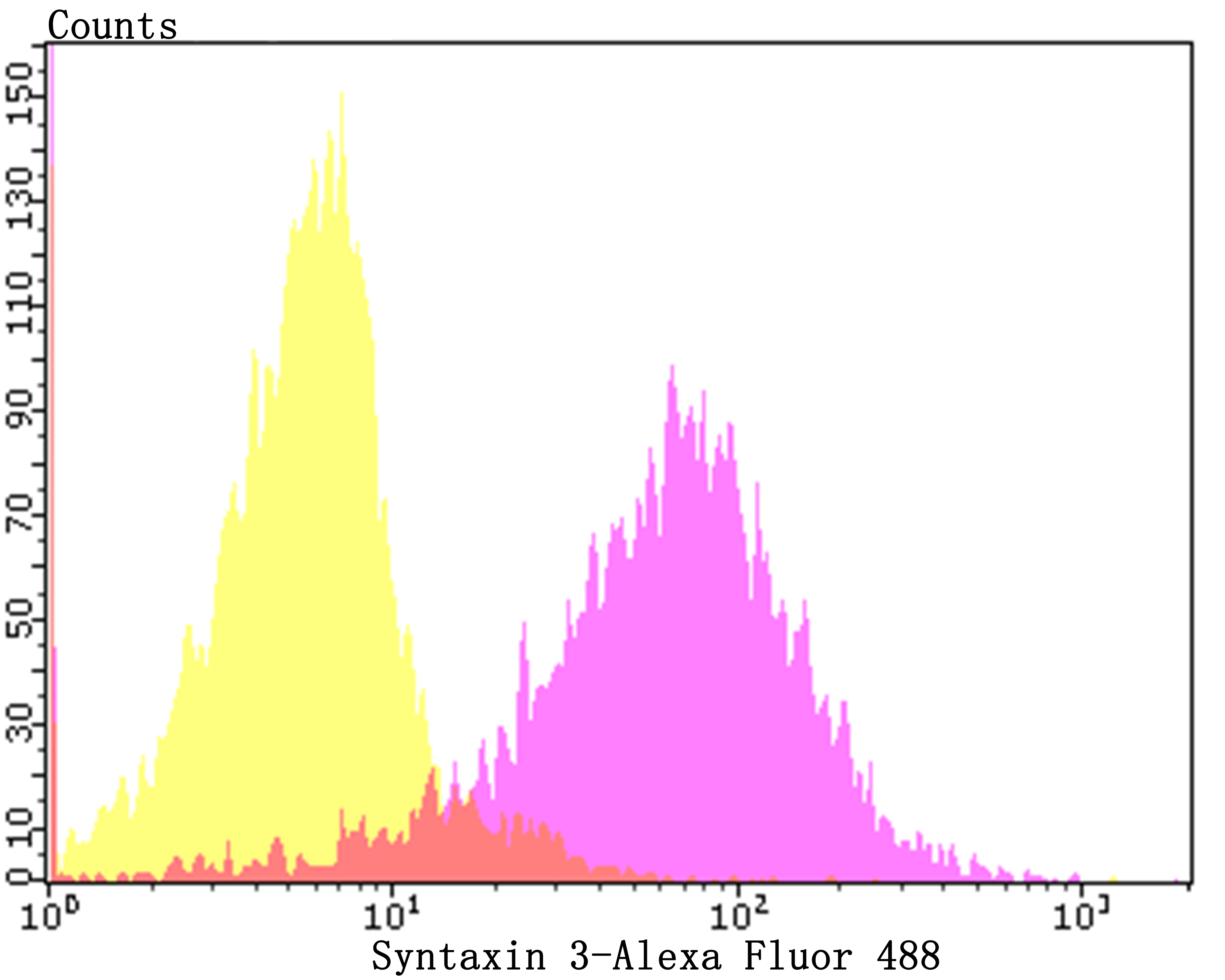
Fig6: Flow cytometric analysis of 293T cells with Syntaxin 3 antibody at 1/100 dilution (purple) compared with an unlabelled control (cells without incubation with primary antibody; yellow). Alexa Fluor 488-conjugated goat anti-rabbit IgG was used as the secondary antibody.Application
Fig6: Flow cytometric analysis of 293T cells with Syntaxin 3 antibody at 1/100 dilution (purple) compared with an unlabelled control (cells without incubation with primary antibody; yellow). Alexa Fluor 488-conjugated goat anti-rabbit IgG was used as the secondary antibody.Application
Fig6: Flow cytometric analysis of 293T cells with Syntaxin 3 antibody at 1/100 dilution (purple) compared with an unlabelled control (cells without incubation with primary antibody; yellow). Alexa Fluor 488-conjugated goat anti-rabbit IgG was used as the secondary antibody.Application
Fig6: Flow cytometric analysis of 293T cells with Syntaxin 3 antibody at 1/100 dilution (purple) compared with an unlabelled control (cells without incubation with primary antibody; yellow). Alexa Fluor 488-conjugated goat anti-rabbit IgG was used as the secondary antibody.Application
Fig6: Flow cytometric analysis of 293T cells with Syntaxin 3 antibody at 1/100 dilution (purple) compared with an unlabelled control (cells without incubation with primary antibody; yellow). Alexa Fluor 488-conjugated goat anti-rabbit IgG was used as the secondary antibody.Application
Fig6: Flow cytometric analysis of 293T cells with Syntaxin 3 antibody at 1/100 dilution (purple) compared with an unlabelled control (cells without incubation with primary antibody; yellow). Alexa Fluor 488-conjugated goat anti-rabbit IgG was used as the secondary antibody.| Product Name | Syntaxin 3 [JE40-96] |
|---|---|
| Antibody Type | Primary Antibodies |
| Antigen Alias | FLJ30906 antibody Stx3 antibody STX3_HUMAN antibody STX3A antibody Syntaxin 3A antibody Syntaxin-3 antibody |
| Product description | Correct vesicular transport is essential to the survival of eukaryotic cells. This process is determined by specific pairing of vesicle-associated SNAREs (v-SNAREs) with those on the target membrane (t-SNAREs). This complex then recruits soluble NSF attachment proteins (SNAPs) and N-ethylmaleimide-sensitive factor (NSF) to form the highly stable SNAP receptor (SNARE) complex. The formation of a SNARE complex pulls the vesicle and target membrane together and may provide the energy to drive fusion of the lipid bilayers. Syntaxins, a family of proteins involved in the fusion of synaptic vesicles with the plasma membrane, display broad tissue distribution and contain carboxy-terminal hydrophobic domains that direct themselves to their respective intracellular compartments. Syntaxin 3 localizes to the apical plasma membrane and is involved in membrane fusion of apical trafficking pathways. Syntaxin 3 is a key factor in the growth of neurites, and it also functions as a direct target for arachidonic acid. Human Syntaxin 3 has two forms: Syntaxin 3A and 3B, while the mouse version has four forms: 3A, 3B, 3C, and 3D. |
| Immunogen | Recombinant protein corresponding to cytoplasmic region of human Syntaxin 3. |
| Clonality | Monoclonal |
|---|---|
| Isotype | IgG |
| Host Species | Recombinant rabbit |
| Tested Applications | WBIHCFC |
| WB:1:500-1:2,000 IHC:1:50-1:200 FC:1:50-1:100 |
|
| Species Reactivity | HumanMouseRat |
| Concentration | 1mg/ml |
| Alternative Names | FLJ30906 antibody Stx3 antibody STX3_HUMAN antibody STX3A antibody Syntaxin 3A antibody Syntaxin-3 antibody |
|---|---|
| Molecular Weight(MW) | 33 kDa |
| Cellular Localization | Membrane. |
Application
Fig6: Flow cytometric analysis of 293T cells with Syntaxin 3 antibody at 1/100 dilution (purple) compared with an unlabelled control (cells without incubation with primary antibody; yellow). Alexa Fluor 488-conjugated goat anti-rabbit IgG was used as the secondary antibody.
Application
Fig6: Flow cytometric analysis of 293T cells with Syntaxin 3 antibody at 1/100 dilution (purple) compared with an unlabelled control (cells without incubation with primary antibody; yellow). Alexa Fluor 488-conjugated goat anti-rabbit IgG was used as the secondary antibody.
Application
Fig6: Flow cytometric analysis of 293T cells with Syntaxin 3 antibody at 1/100 dilution (purple) compared with an unlabelled control (cells without incubation with primary antibody; yellow). Alexa Fluor 488-conjugated goat anti-rabbit IgG was used as the secondary antibody.
Application
Fig6: Flow cytometric analysis of 293T cells with Syntaxin 3 antibody at 1/100 dilution (purple) compared with an unlabelled control (cells without incubation with primary antibody; yellow). Alexa Fluor 488-conjugated goat anti-rabbit IgG was used as the secondary antibody.
Application
Fig6: Flow cytometric analysis of 293T cells with Syntaxin 3 antibody at 1/100 dilution (purple) compared with an unlabelled control (cells without incubation with primary antibody; yellow). Alexa Fluor 488-conjugated goat anti-rabbit IgG was used as the secondary antibody.
Application
Fig6: Flow cytometric analysis of 293T cells with Syntaxin 3 antibody at 1/100 dilution (purple) compared with an unlabelled control (cells without incubation with primary antibody; yellow). Alexa Fluor 488-conjugated goat anti-rabbit IgG was used as the secondary antibody.| Positive Control | SH-SY-5Y, human kidney tissue, rat kidney tissue, human prostate cancer tissue, mouse pancreas tissue. |
|---|---|
| Application Notes | WB:1:500-1:2,000 IHC:1:50-1:200 FC:1:50-1:100 |
| Form | Liquid |
|---|---|
| Storage Instructions | Store at +4℃ after thawing. Aliquot store at -20℃. Avoid repeated freeze / thaw cycles. |
| Storage Buffer | 1*TBS (pH7.4), 1%BSA, 40%Glycerol. Preservative: 0.05% Sodium Azide. |


