Application
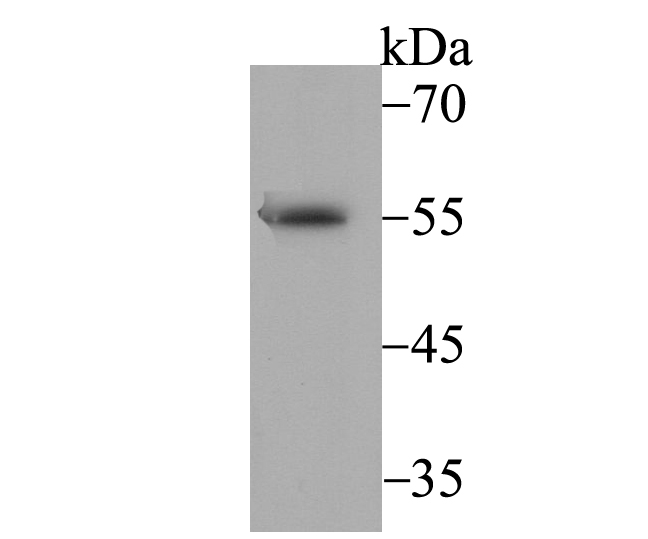
Fig1: Western blot analysis of BAF57 on SH-SY-5Y cell lysate using anti-BAF57 antibody at 1/1,000 dilution.Application
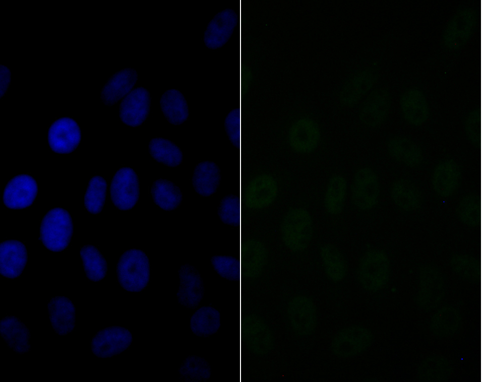
Fig2: ICC staining BAF57 in HepG2 cells (green). The nuclear counter stain is DAPI (blue). Cells were fixed in paraformaldehyde, permeabilised with 0.25% Triton X100/PBS.Application
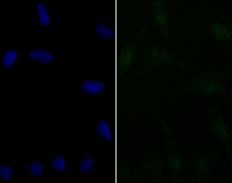
Fig3: ICC staining BAF57 in SH-SY-5Y cells (green). The nuclear counter stain is DAPI (blue). Cells were fixed in paraformaldehyde, permeabilised with 0.25% Triton X100/PBS.Application
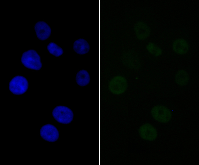
Fig4: ICC staining BAF57 in SK-Br-3 cells (green). The nuclear counter stain is DAPI (blue). Cells were fixed in paraformaldehyde, permeabilised with 0.25% Triton X100/PBS.Application
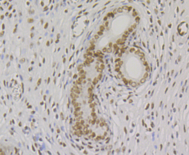
Fig5: Immunohistochemical analysis of paraffin-embedded rat uterus tissue using anti-BAF57 antibody. Counter stained with hematoxylin.Application
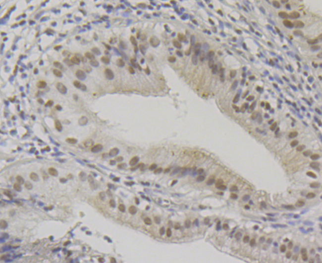
Fig6: Immunohistochemical analysis of paraffin-embedded human gallbladder tissue using anti-BAF57 antibody. Counter stained with hematoxylin.Application
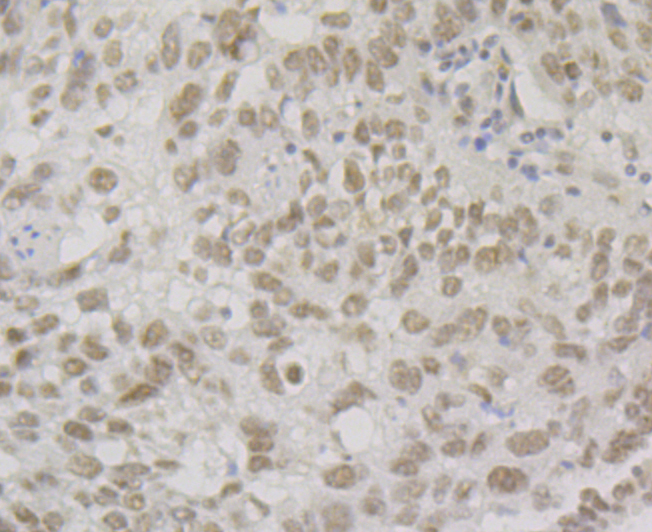
Fig7: Immunohistochemical analysis of paraffin-embedded human lung cancer tissue using anti-BAF57/SMARCE1 antibody. Counter stained with hematoxylin.Application
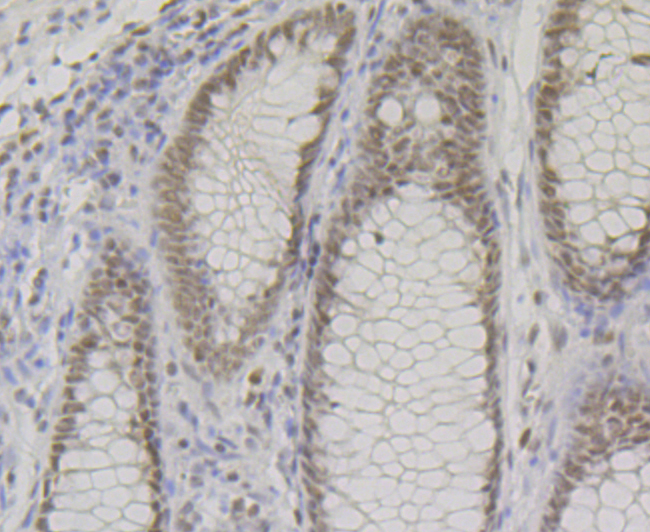
Fig8: Immunohistochemical analysis of paraffin-embedded human colon tissue using anti-BAF57 antibody. Counter stained with hematoxylin.| Product Name | BAF57 Recombinant Rabbit Monoclonal Antibody |
|---|---|
| Antibody Type | Primary Antibodies |
| Product description | The SWI/SNF complex is involved in the activation of transcription via the remodeling of nucleosome structure in an ATP-dependent manner. Brm (also designated SNF1 or SNF2α) and Brg-1 (also designated SNF2 or SNF2β) are the ATPase subunits of the mammalian SWI/SNF complex. Brm, Brg-1, Ini1 (integrase interactor 1, also designated SNF5), BAF155 (also designated SRG3) and BAF170 are thought to comprise the functional core of the SWI/SNF complex. In higher eukaryotes, BAF57 is also a critical component of the SWI/ SNF complex. BAF57 contains a high-mobility-group (HMG) domain adjacent to a kinesin-like region and is a DNA-binding subunit of the SWI/SNF complex. The human BAF57 gene maps within the q12-25 region of chromosome 17, a gene-rich area implicated in breast and ovarian cancers. |
| Immunogen | Recombinant protein within human BAF57 aa 1-200. |
| Clonality | Monoclonal |
|---|---|
| Isotype | IgG |
| Host Species | Recombinant rabbit |
| Tested Applications | ICCIFIHCIPWB |
| WB:1:500-1:2,000 ICC/IF:1:50-1:200 IHC:1:50-1:200 IP:1:50-1:200 |
|
| Species Reactivity | HumanMouseRat |
| Concentration | 1mg/ml |
| Purification | Protein A |
| Alternative Names | BAF57 antibody BRG1 associated factor 57 antibody BRG1-associated factor 57 antibody Chromatin remodeling complex BRG1 associated factor 57 antibody FLJ35648 antibody SMARCE 1 antibody SMARCE1 antibody SMCE1_HUMAN antibody SWI/SNF related matrix associated actin dependent regulator of chromatin e1 antibody SWI/SNF related matrix associated actin dependent regulator of chromatin subfamily e member 1 antibody SWI/SNF-related matrix-associated actin-dependent regulator of chromatin subfamily E member 1 antibody |
|---|---|
| Molecular Weight(MW) | 47 kDa |
| Cellular Localization | Nucleus. |
Application
Fig1: Western blot analysis of BAF57 on SH-SY-5Y cell lysate using anti-BAF57 antibody at 1/1,000 dilution.
Application
Fig2: ICC staining BAF57 in HepG2 cells (green). The nuclear counter stain is DAPI (blue). Cells were fixed in paraformaldehyde, permeabilised with 0.25% Triton X100/PBS.
Application
Fig3: ICC staining BAF57 in SH-SY-5Y cells (green). The nuclear counter stain is DAPI (blue). Cells were fixed in paraformaldehyde, permeabilised with 0.25% Triton X100/PBS.
Application
Fig4: ICC staining BAF57 in SK-Br-3 cells (green). The nuclear counter stain is DAPI (blue). Cells were fixed in paraformaldehyde, permeabilised with 0.25% Triton X100/PBS.
Application
Fig5: Immunohistochemical analysis of paraffin-embedded rat uterus tissue using anti-BAF57 antibody. Counter stained with hematoxylin.
Application
Fig6: Immunohistochemical analysis of paraffin-embedded human gallbladder tissue using anti-BAF57 antibody. Counter stained with hematoxylin.
Application
Fig7: Immunohistochemical analysis of paraffin-embedded human lung cancer tissue using anti-BAF57/SMARCE1 antibody. Counter stained with hematoxylin.
Application
Fig8: Immunohistochemical analysis of paraffin-embedded human colon tissue using anti-BAF57 antibody. Counter stained with hematoxylin.| Positive Control | SH-SY-5Y, HepG2, SH-SY-5Y, SK-Br-3, rat uterus tissue, human gallbladder tissue, human lung cancer tissue, human colon tissue. |
|---|---|
| Application Notes | WB:1:500-1:2,000 ICC/IF:1:50-1:200 IHC:1:50-1:200 IP:1:50-1:200 |
| Form | Liquid |
|---|---|
| Storage Instructions | Store at +4℃ after thawing. Aliquot store at -20℃. Avoid repeated freeze / thaw cycles. |
| Storage Buffer | 1*TBS (pH7.4), 1%BSA, 40%Glycerol. Preservative: 0.05% Sodium Azide. |


