Application
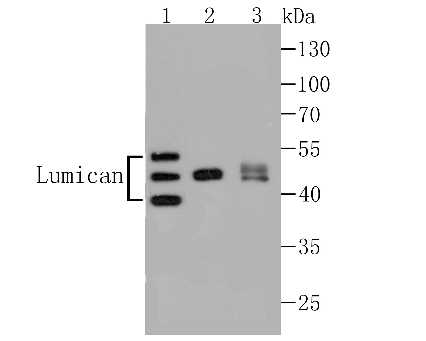
Fig1: Western blot analysis of Lumican on different lysates using anti-Lumican antibody at 1/1,000 dilution. Positive control: Lane 1: Jurkat Lane 2: 293T Lane 3: Human skinApplication
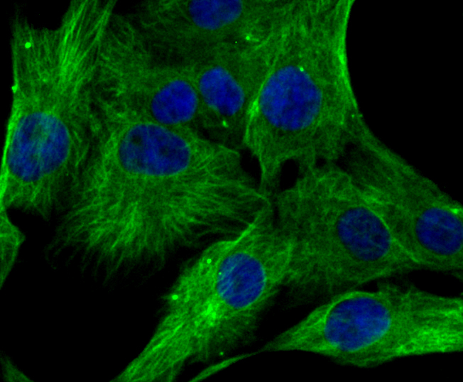
Fig2: ICC staining Lumican in MG-63 cells (green). The nuclear counter stain is DAPI (blue). Cells were fixed in paraformaldehyde, permeabilised with 0.25% Triton X100/PBS.Application
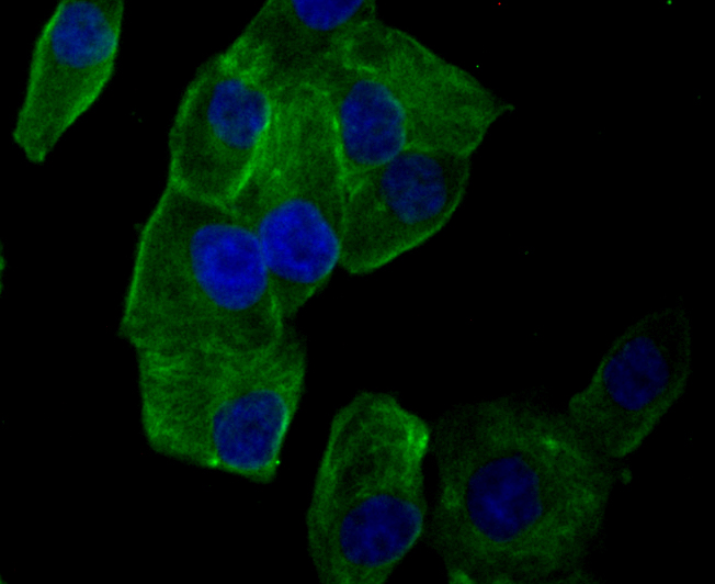
Fig3: ICC staining Lumican in PANC-1 cells (green). The nuclear counter stain is DAPI (blue). Cells were fixed in paraformaldehyde, permeabilised with 0.25% Triton X100/PBS.Application
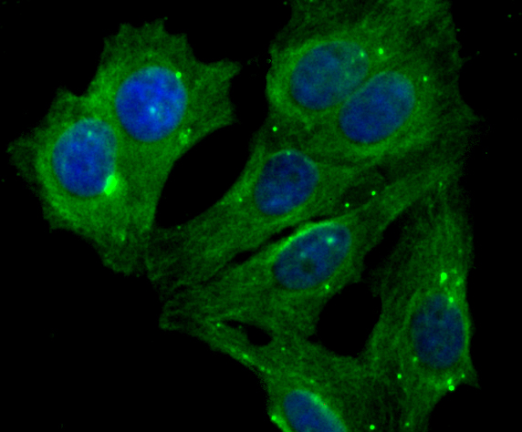
Fig4: ICC staining Lumican in MCF-7 cells (green). The nuclear counter stain is DAPI (blue). Cells were fixed in paraformaldehyde, permeabilised with 0.25% Triton X100/PBS.Application
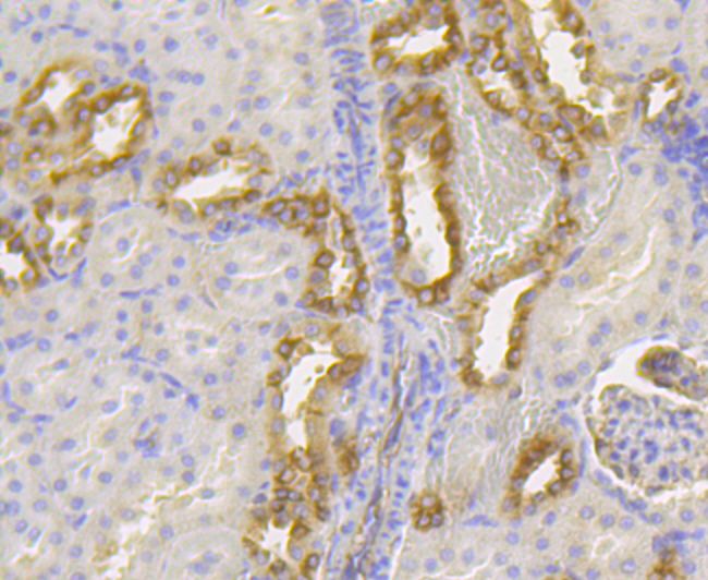
Fig5: Immunohistochemical analysis of paraffin-embedded rat kidney tissue using anti-Lumican antibody. Counter stained with hematoxylin.Application
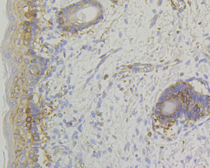
Fig6: Immunohistochemical analysis of paraffin-embedded human skin tissue using anti-Lumican antibody. Counter stained with hematoxylin.Application
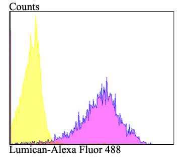
Fig7: Flow cytometric analysis of 293T cells with Lumican antibody at 1/100 dilution (purple) compared with an unlabelled control (cells without incubation with primary antibody; yellow). Alexa Fluor 488-conjugated goat anti-rabbit IgG was used as the secondary antibody.| Product Name | Lumican [JE11-45] |
|---|---|
| Antibody Type | Primary Antibodies |
| Product description | Lumican, a member of the small leucine-rich proteoglycans (SLRPs) family of proteins, is a keratan sulfate proteoglycan present in large quantities in the corneal stroma and in interstitial collagenous matrices of the heart, aorta, skeletal muscle, skin, and intervertebral discs. During collagen fibrillogenesis, lumican regulates the assembly of collagens into higher order fibrils, limiting the growth of fibrils in diameter. Lumican is present in the extracellular matrix of human articular cartilage at all ages, with a far greater abundance in the adult. The gene encoding human lumican maps to chromosome 12q21.3-q22. |
| Immunogen | Recombinant protein within human Lumican aa 50-250. |
| Clonality | Monoclonal |
|---|---|
| Isotype | IgG |
| Host Species | Recombinant rabbit |
| Tested Applications | WB,ICC,IHC,FC |
| WB:1:500-1:2,000 ICC:1:50-1:200 IHC:1:50-1:200 FC:1:50-1:100 |
|
| Species Reactivity | Human,Mouse,Rat |
| Concentration | 1mg/ml |
| Purification | Unpurified |
| Alternative Names | Keratan sulfate proteoglycan lumican antibody KSPG lumican antibody LDC antibody LUM antibody LUM_HUMAN antibody Lumican antibody Lumican precursor antibody SLRR2D antibody |
|---|---|
| Molecular Weight(MW) | 38 kDa |
| Cellular Localization | Extracellular matrix. Secreted. |
| SwissProt ID | P51884 |
|---|
Application
Fig1: Western blot analysis of Lumican on different lysates using anti-Lumican antibody at 1/1,000 dilution. Positive control: Lane 1: Jurkat Lane 2: 293T Lane 3: Human skin
Application
Fig2: ICC staining Lumican in MG-63 cells (green). The nuclear counter stain is DAPI (blue). Cells were fixed in paraformaldehyde, permeabilised with 0.25% Triton X100/PBS.
Application
Fig3: ICC staining Lumican in PANC-1 cells (green). The nuclear counter stain is DAPI (blue). Cells were fixed in paraformaldehyde, permeabilised with 0.25% Triton X100/PBS.
Application
Fig4: ICC staining Lumican in MCF-7 cells (green). The nuclear counter stain is DAPI (blue). Cells were fixed in paraformaldehyde, permeabilised with 0.25% Triton X100/PBS.
Application
Fig5: Immunohistochemical analysis of paraffin-embedded rat kidney tissue using anti-Lumican antibody. Counter stained with hematoxylin.
Application
Fig6: Immunohistochemical analysis of paraffin-embedded human skin tissue using anti-Lumican antibody. Counter stained with hematoxylin.
Application
Fig7: Flow cytometric analysis of 293T cells with Lumican antibody at 1/100 dilution (purple) compared with an unlabelled control (cells without incubation with primary antibody; yellow). Alexa Fluor 488-conjugated goat anti-rabbit IgG was used as the secondary antibody.| Positive Control | Jurkat, 293T, Human skin tissue, MCF-7, MG-63, PANC-1, rat kidney tissue, human skin tissue. |
|---|---|
| Application Notes | WB:1:500-1:2,000 ICC:1:50-1:200 IHC:1:50-1:200 FC:1:50-1:100 |
| Form | Liquid |
|---|---|
| Storage Instructions | Store at +4℃ after thawing. Aliquot store at -20℃. Avoid repeated freeze / thaw cycles. |
| Storage Buffer | 1*TBS (pH7.4), 1%BSA, 40%Glycerol. Preservative: 0.05% Sodium Azide. |


