Application
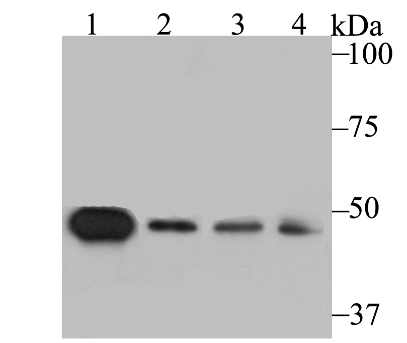
Fig1: Western blot analysis of SFRP4 on different lysates using anti-SFRP4 antibody at 1/1,000 dilution. Positive control: Lane 1: Mouse testis Lane 2: A549 Lane 3: HepG2 Lane 4: SH-SY-5YApplication
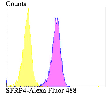
Fig2: Flow cytometric analysis of A549 cells with SFRP4 antibody at 1/100 dilution (purple) compared with an unlabelled control (cells without incubation with primary antibody; yellow). Alexa Fluor 488-conjugated goat anti-rabbit IgG was used as the secondary antibody.| Product Name | SFRP4 [JE41-41] |
|---|---|
| Antibody Type | Primary Antibodies |
| Antigen Alias | Frizzled protein antibody Frizzled protein human endometrium antibody FRP 4 antibody FRP4 antibody FrpHE antibody human endometrium antibody PYL antibody Secreted frizzled-related protein 4 antibody sFRP-4 antibody Sfrp4 antibody SFRP4_HUMAN antibody |
| Product description | Soluble frizzled-related proteins (sFRPS) function as modulators of Wnt signaling through direct interaction with Wnts. They have a role in regulating cell growth and differentiation in specific cell types. SFRP4 plays a role in bone morphogenesis. May also act as a regulator of adult uterine morphology and function. May also increase apoptosis during ovulation possibly through modulation of FZ1/FZ4/WNT4 signaling. Has phosphaturic effects by specifically inhibiting sodium-dependent phosphate uptake. |
| Immunogen | Full length recombinant protein of human SFRP4. |
| Clonality | Monoclonal |
|---|---|
| Isotype | IgG |
| Host Species | Recombinant rabbit |
| Tested Applications | WBFC |
| WB:1:500-1:2,000 FC:1:50-1:100 |
|
| Species Reactivity | HumanMouseRat |
| Concentration | 1mg/ml |
| Alternative Names | Frizzled protein antibody Frizzled protein human endometrium antibody FRP 4 antibody FRP4 antibody FrpHE antibody human endometrium antibody PYL antibody Secreted frizzled-related protein 4 antibody sFRP-4 antibody Sfrp4 antibody SFRP4_HUMAN antibody |
|---|---|
| Molecular Weight(MW) | 40 kDa |
| Cellular Localization | Secreted. |
Application
Fig1: Western blot analysis of SFRP4 on different lysates using anti-SFRP4 antibody at 1/1,000 dilution. Positive control: Lane 1: Mouse testis Lane 2: A549 Lane 3: HepG2 Lane 4: SH-SY-5Y
Application
Fig2: Flow cytometric analysis of A549 cells with SFRP4 antibody at 1/100 dilution (purple) compared with an unlabelled control (cells without incubation with primary antibody; yellow). Alexa Fluor 488-conjugated goat anti-rabbit IgG was used as the secondary antibody.| Positive Control | Mouse testis tissue, A549, HepG2, SH-SY-5Y. |
|---|---|
| Application Notes | WB:1:500-1:2,000 FC:1:50-1:100 |
| Form | Liquid |
|---|---|
| Storage Instructions | Store at +4℃ after thawing. Aliquot store at -20℃. Avoid repeated freeze / thaw cycles. |
| Storage Buffer | 1*TBS (pH7.4), 1%BSA, 40%Glycerol. Preservative: 0.05% Sodium Azide. |


