Application
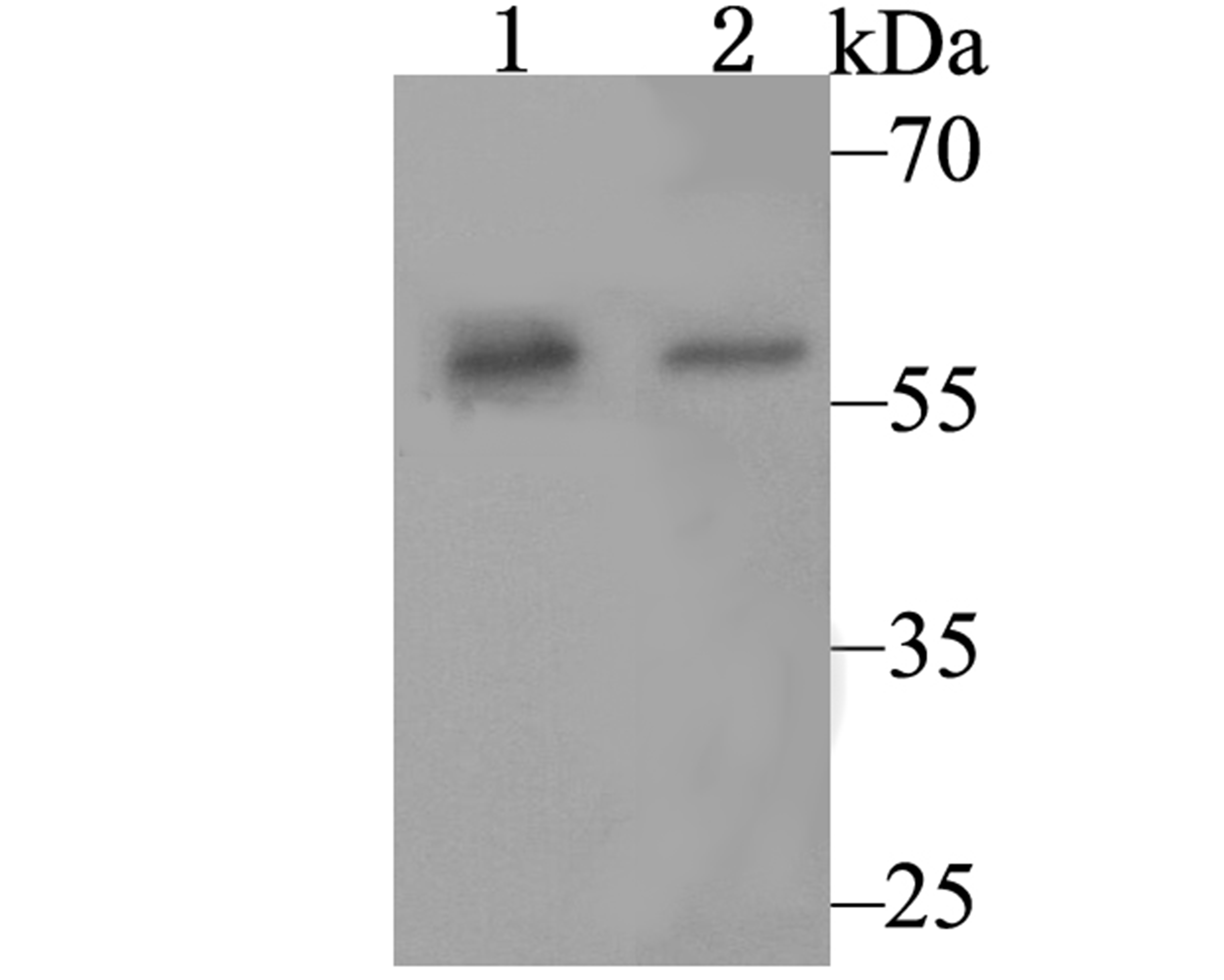
Fig1: Western blot analysis of SRP54 on different lysates. Proteins were transferred to a PVDF membrane and blocked with 5% BSA in PBS for 1 hour at room temperature. The primary antibody was used at a 1:500 dilution in 5% BSA at room temperature for 2 hours. Goat Anti-Rabbit IgG - HRP Secondary Antibody (HA1001) at 1:5,000 dilution was used for 1 hour at room temperature. Positive control: Lane 1: HepG2 Lane 2: MCF-7Application
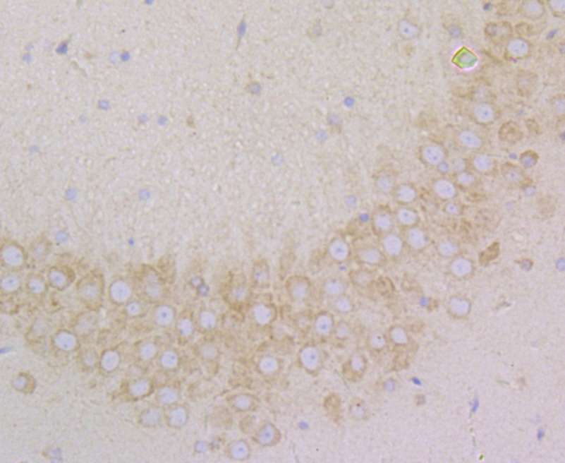
Fig2: Immunohistochemical analysis of paraffin-embedded rat brain tissue using anti-SRP54 antibody. The section was pre-treated using heat mediated antigen retrieval with sodium citrate buffer (pH 6.0) for 20 minutes. The tissues were blocked in 5% BSA for 30 minutes at room temperature, washed with ddH2O and PBS, and then probed with the antibody (ET7109-32) at 1/200 dilution, for 30 minutes at room temperature and detected using an HRP conjugated compact polymer system. DAB was used as the chrogen. Counter stained with hematoxylin and mounted with DPX.Application
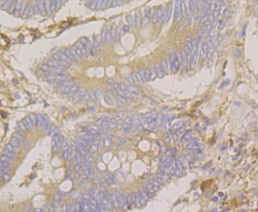
Fig3: Immunohistochemical analysis of paraffin-embedded human breast cancer tissue using anti-SRP54 antibody. The section was pre-treated using heat mediated antigen retrieval with sodium citrate buffer (pH 6.0) for 20 minutes. The tissues were blocked in 5% BSA for 30 minutes at room temperature, washed with ddH2O and PBS, and then probed with the antibody (ET7109-32) at 1/200 dilution, for 30 minutes at room temperature and detected using an HRP conjugated compact polymer system. DAB was used as the chrogen. Counter stained with hematoxylin and mounted with DPX.Application
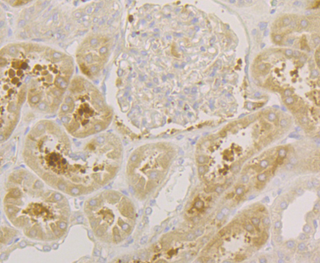
Fig4: Immunohistochemical analysis of paraffin-embedded human kidney tissue using anti-SRP54 antibody. The section was pre-treated using heat mediated antigen retrieval with sodium citrate buffer (pH 6.0) for 20 minutes. The tissues were blocked in 5% BSA for 30 minutes at room temperature, washed with ddH2O and PBS, and then probed with the antibody (ET7109-32) at 1/200 dilution, for 30 minutes at room temperature and detected using an HRP conjugated compact polymer system. DAB was used as the chrogen. Counter stained with hematoxylin and mounted with DPX.Application
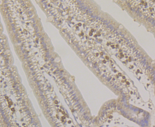
Fig5: Immunohistochemical analysis of paraffin-embedded mouse colon tissue using anti-SRP54 antibody. The section was pre-treated using heat mediated antigen retrieval with sodium citrate buffer (pH 6.0) for 20 minutes. The tissues were blocked in 5% BSA for 30 minutes at room temperature, washed with ddH2O and PBS, and then probed with the antibody (ET7109-32) at 1/200 dilution, for 30 minutes at room temperature and detected using an HRP conjugated compact polymer system. DAB was used as the chrogen. Counter stained with hematoxylin and mounted with DPX.Application
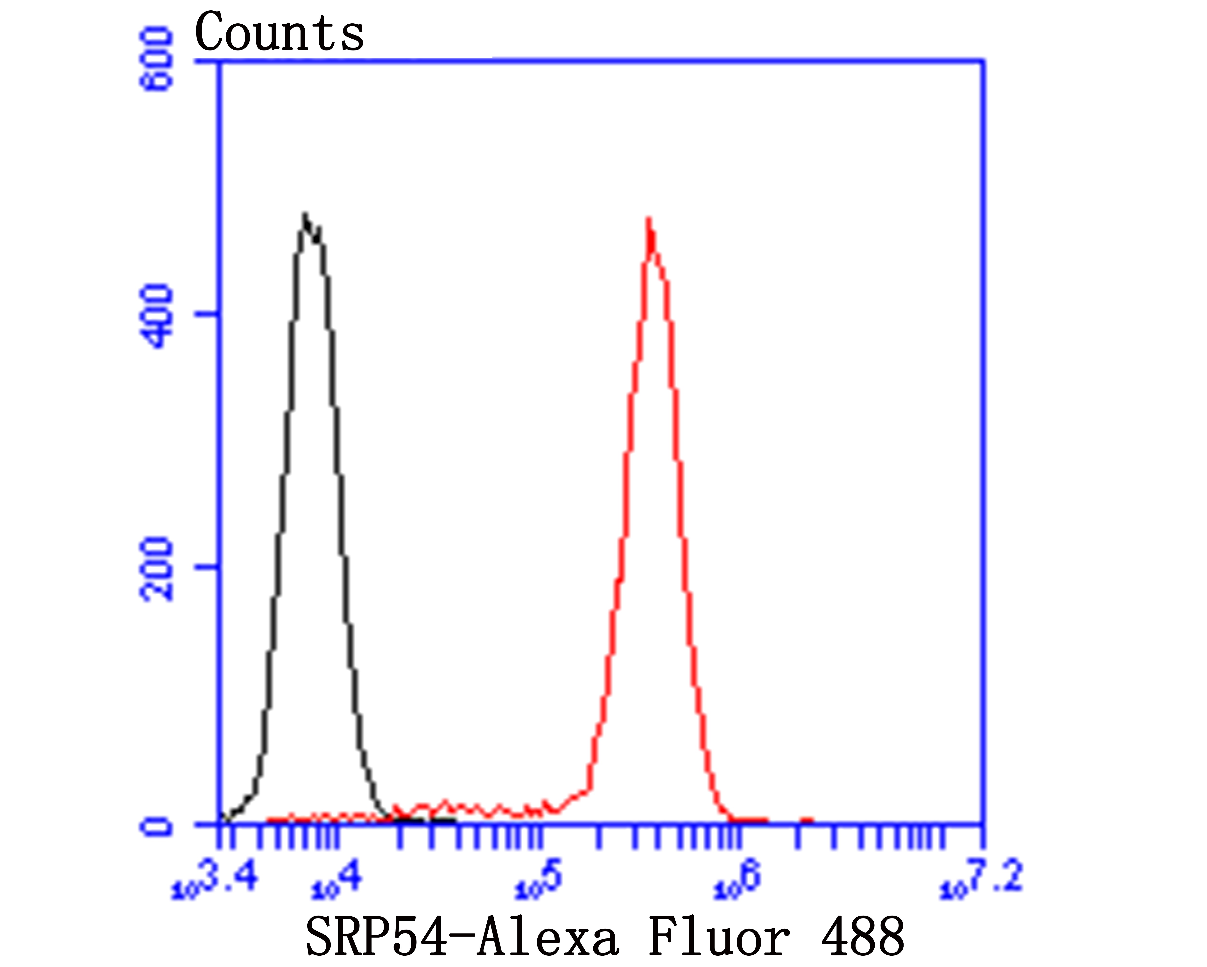
Fig6: Flow cytometric analysis of SRP54 was done on HepG2 cells. The cells were fixed, permeabilized and stained with ClC-5 antibody at 1/100 dilution (red) compared with an unlabelled control (cells without incubation with primary antibody; black). After incubation of the primary antibody on room temperature for an hour, the cells was stained with a Alexa Fluor™ 488-conjugated goat anti-rabbit IgG Secondary antibody at 1/500 dilution for 30 minutes.| Product Name | SRP54 [JE43-25] |
|---|---|
| Antibody Type | Primary Antibodies |
| Product description | SRP54, a component of the signal recognition particle (SRP), recognizes the signal peptide of secretory proteins and interacts with the signal recognition particle receptor to target the ribosome and the associated nascent chain to the endoplasmic reticulum. By functional analysis showed that SRP54 binds to SRP RNA via the M-domain, but only in the presence of RNA-bound SRP19, and that it associates with the signal peptide of nascent polypeptide chains. RNA interaction requires the presence of a loop in the C-terminal M-domain. Signal peptide recognition most likely involves methionine-rich loops. During signal peptide recognition, SRP54 is positioned at the exit site close to ribosomal proteins L23a (602326) and L35. When SRP54 contacts the signal recognition particle receptor (182180), SRP54 is rearranged such that it is no longer close to L23a. This repositioning may allow the translocon to dock with the ribosome, leading to insertion of the signal peptide into the translocation channel. |
| Immunogen | Recombinant protein within human SRP54 aa 1-200. |
| Clonality | Monoclonal |
|---|---|
| Isotype | IgG |
| Host Species | Recombinant rabbit |
| Tested Applications | WB,IHC,FC,IP |
| WB:1:500IP:1:10-1:50IHC:1:50-1:200FC:1:50-1:100 | |
| Species Reactivity | Human,Mouse,Rat |
| Concentration | 1mg/ml |
| Purification | Unpurified |
| Alternative Names | Signal recognition particle 54 kDa protein antibody Signal recognition particle 54kDa antibody SRP 54 antibody SRP54 antibody SRP54_HUMAN antibody |
|---|---|
| Molecular Weight(MW) | 56 kDa |
| Cellular Localization | Cytoplasm, Nucleus. |
Application
Fig1: Western blot analysis of SRP54 on different lysates. Proteins were transferred to a PVDF membrane and blocked with 5% BSA in PBS for 1 hour at room temperature. The primary antibody was used at a 1:500 dilution in 5% BSA at room temperature for 2 hours. Goat Anti-Rabbit IgG - HRP Secondary Antibody (HA1001) at 1:5,000 dilution was used for 1 hour at room temperature. Positive control: Lane 1: HepG2 Lane 2: MCF-7
Application
Fig2: Immunohistochemical analysis of paraffin-embedded rat brain tissue using anti-SRP54 antibody. The section was pre-treated using heat mediated antigen retrieval with sodium citrate buffer (pH 6.0) for 20 minutes. The tissues were blocked in 5% BSA for 30 minutes at room temperature, washed with ddH2O and PBS, and then probed with the antibody (ET7109-32) at 1/200 dilution, for 30 minutes at room temperature and detected using an HRP conjugated compact polymer system. DAB was used as the chrogen. Counter stained with hematoxylin and mounted with DPX.
Application
Fig3: Immunohistochemical analysis of paraffin-embedded human breast cancer tissue using anti-SRP54 antibody. The section was pre-treated using heat mediated antigen retrieval with sodium citrate buffer (pH 6.0) for 20 minutes. The tissues were blocked in 5% BSA for 30 minutes at room temperature, washed with ddH2O and PBS, and then probed with the antibody (ET7109-32) at 1/200 dilution, for 30 minutes at room temperature and detected using an HRP conjugated compact polymer system. DAB was used as the chrogen. Counter stained with hematoxylin and mounted with DPX.
Application
Fig4: Immunohistochemical analysis of paraffin-embedded human kidney tissue using anti-SRP54 antibody. The section was pre-treated using heat mediated antigen retrieval with sodium citrate buffer (pH 6.0) for 20 minutes. The tissues were blocked in 5% BSA for 30 minutes at room temperature, washed with ddH2O and PBS, and then probed with the antibody (ET7109-32) at 1/200 dilution, for 30 minutes at room temperature and detected using an HRP conjugated compact polymer system. DAB was used as the chrogen. Counter stained with hematoxylin and mounted with DPX.
Application
Fig5: Immunohistochemical analysis of paraffin-embedded mouse colon tissue using anti-SRP54 antibody. The section was pre-treated using heat mediated antigen retrieval with sodium citrate buffer (pH 6.0) for 20 minutes. The tissues were blocked in 5% BSA for 30 minutes at room temperature, washed with ddH2O and PBS, and then probed with the antibody (ET7109-32) at 1/200 dilution, for 30 minutes at room temperature and detected using an HRP conjugated compact polymer system. DAB was used as the chrogen. Counter stained with hematoxylin and mounted with DPX.
Application
Fig6: Flow cytometric analysis of SRP54 was done on HepG2 cells. The cells were fixed, permeabilized and stained with ClC-5 antibody at 1/100 dilution (red) compared with an unlabelled control (cells without incubation with primary antibody; black). After incubation of the primary antibody on room temperature for an hour, the cells was stained with a Alexa Fluor™ 488-conjugated goat anti-rabbit IgG Secondary antibody at 1/500 dilution for 30 minutes.| Positive Control | HepG2, rat brain tissue, human colon cancer tissue, human kidney tissue, mouse small intestine tissue. |
|---|---|
| Application Notes | WB:1:500IP:1:10-1:50IHC:1:50-1:200FC:1:50-1:100 |
| Form | Liquid |
|---|---|
| Storage Instructions | Store at +4℃ after thawing. Aliquot store at -20℃. Avoid repeated freeze / thaw cycles. |
| Storage Buffer | 1*TBS (pH7.4), 1%BSA, 50%Glycerol. Preservative: 0.05% Sodium Azide. |


