Application
Fig5: Flow cytometric analysis of FYB was done on HepG2 cells. The cells were fixed, permeabilized and stained with FYB antibody at 1/100 dilution (red) compared with an unlabelled control (cells without incubation with primary antibody; black). After incubation of the primary antibody on room temperature for 1 hour, the cells was stained with a Alexa Fluor™ 488-conjugated goat anti-rabbit IgG Secondary antibody at 1/500 dilution for 30 minutes.Application
Fig5: Flow cytometric analysis of FYB was done on HepG2 cells. The cells were fixed, permeabilized and stained with FYB antibody at 1/100 dilution (red) compared with an unlabelled control (cells without incubation with primary antibody; black). After incubation of the primary antibody on room temperature for 1 hour, the cells was stained with a Alexa Fluor™ 488-conjugated goat anti-rabbit IgG Secondary antibody at 1/500 dilution for 30 minutes.Application
Fig5: Flow cytometric analysis of FYB was done on HepG2 cells. The cells were fixed, permeabilized and stained with FYB antibody at 1/100 dilution (red) compared with an unlabelled control (cells without incubation with primary antibody; black). After incubation of the primary antibody on room temperature for 1 hour, the cells was stained with a Alexa Fluor™ 488-conjugated goat anti-rabbit IgG Secondary antibody at 1/500 dilution for 30 minutes.Application
Fig5: Flow cytometric analysis of FYB was done on HepG2 cells. The cells were fixed, permeabilized and stained with FYB antibody at 1/100 dilution (red) compared with an unlabelled control (cells without incubation with primary antibody; black). After incubation of the primary antibody on room temperature for 1 hour, the cells was stained with a Alexa Fluor™ 488-conjugated goat anti-rabbit IgG Secondary antibody at 1/500 dilution for 30 minutes.Application
Fig5: Flow cytometric analysis of FYB was done on HepG2 cells. The cells were fixed, permeabilized and stained with FYB antibody at 1/100 dilution (red) compared with an unlabelled control (cells without incubation with primary antibody; black). After incubation of the primary antibody on room temperature for 1 hour, the cells was stained with a Alexa Fluor™ 488-conjugated goat anti-rabbit IgG Secondary antibody at 1/500 dilution for 30 minutes.| Product Name | FYB [JE44-80] |
|---|---|
| Antibody Type | Primary Antibodies |
| Antigen Alias | ADAP antibody Adhesion and degranulation promoting adaptor protein antibody FYB 120/130 antibody Fyb antibody FYB-120/130 antibody FYB_HUMAN antibody FYN binding protein antibody FYN T binding protein antibody FYN-binding protein antibody FYN-T-binding protein antibody p120/p130 antibody PRO0823 antibody SLAP 130 antibody SLAP-130 antibody SLAP130 antibody SLP 76 associated phosphoprotein antibody SLP-76-associated phosphoprotein antibody SLP76 associated phosphoprotein antibody |
| Product description | Acts as an adapter protein of the FYN and LCP2 signaling cascades in T-cells. Modulates the expression of interleukin-2 (IL-2). Involved in platelet activation. Prevents the degradation of SKAP1 and SKAP2. May play a role in linking T-cell signaling to remodeling of the actin cytoskeleton. Expressed in hematopoietic tissues such as myeloid and T-cells, spleen and thymus. Not expressed in B-cells, nor in non-lymphoid tissues. |
| Immunogen | Recombinant protein within C-terminal human FYB. |
| Clonality | Monoclonal |
|---|---|
| Isotype | IgG |
| Host Species | Recombinant rabbit |
| Tested Applications | WBIHCFC |
| WB:1:500-1:1,000 IHC:1:50-1:200 FC:1:50-1:100 |
|
| Species Reactivity | Human |
| Concentration | 1mg/ml |
| Alternative Names | ADAP antibody Adhesion and degranulation promoting adaptor protein antibody FYB 120/130 antibody Fyb antibody FYB-120/130 antibody FYB_HUMAN antibody FYN binding protein antibody FYN T binding protein antibody FYN-binding protein antibody FYN-T-binding protein antibody p120/p130 antibody PRO0823 antibody SLAP 130 antibody SLAP-130 antibody SLAP130 antibody SLP 76 associated phosphoprotein antibody SLP-76-associated phosphoprotein antibody SLP76 associated phosphoprotein antibody |
|---|---|
| Molecular Weight(MW) | 120 kDa, predicted band size 85 kDa |
| Cellular Localization | Cell junction, Cytoplasm, Nucleus. |
| SwissProt ID | O15117 |
|---|
Application
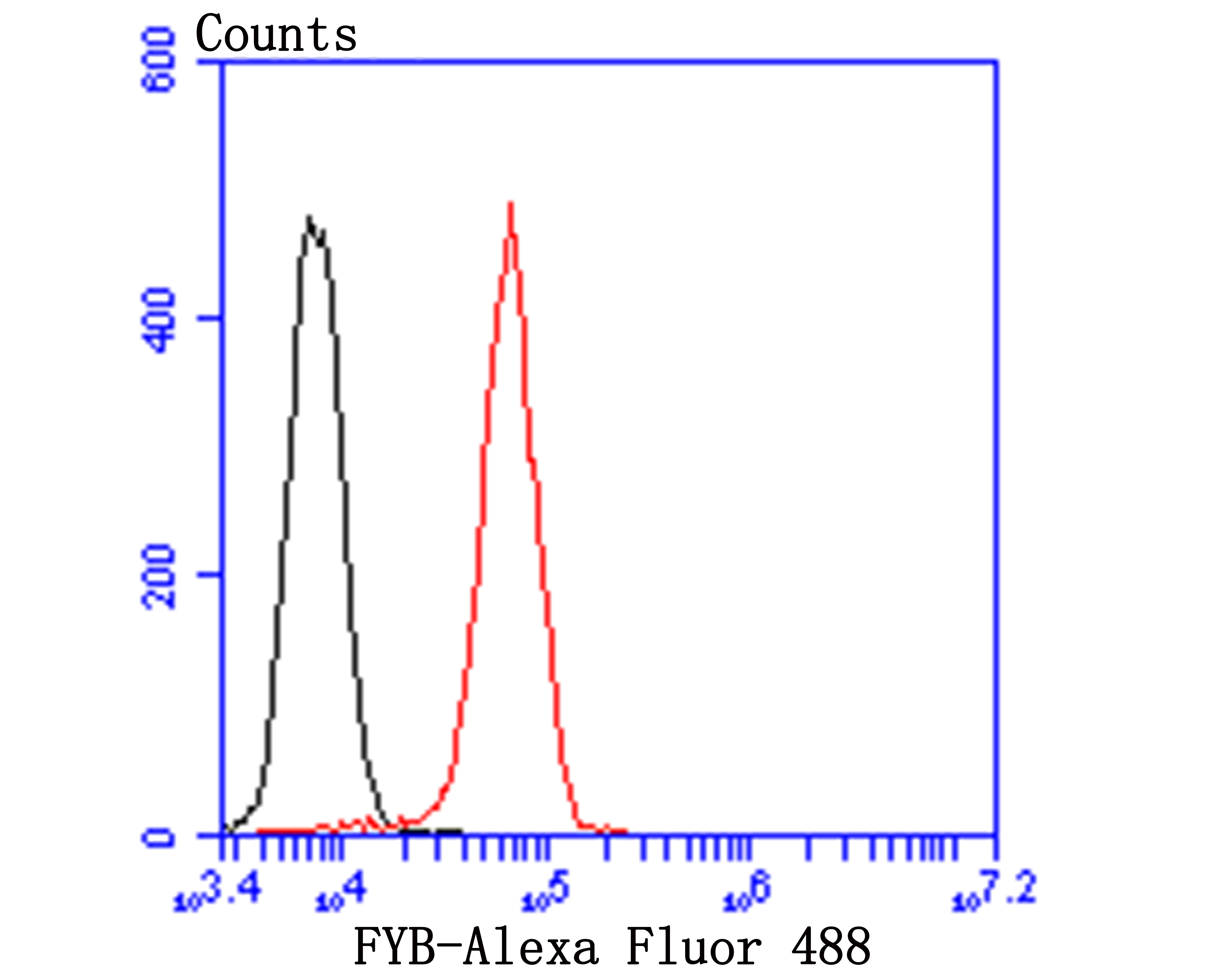
Fig5: Flow cytometric analysis of FYB was done on HepG2 cells. The cells were fixed, permeabilized and stained with FYB antibody at 1/100 dilution (red) compared with an unlabelled control (cells without incubation with primary antibody; black). After incubation of the primary antibody on room temperature for 1 hour, the cells was stained with a Alexa Fluor™ 488-conjugated goat anti-rabbit IgG Secondary antibody at 1/500 dilution for 30 minutes.
Application
Fig5: Flow cytometric analysis of FYB was done on HepG2 cells. The cells were fixed, permeabilized and stained with FYB antibody at 1/100 dilution (red) compared with an unlabelled control (cells without incubation with primary antibody; black). After incubation of the primary antibody on room temperature for 1 hour, the cells was stained with a Alexa Fluor™ 488-conjugated goat anti-rabbit IgG Secondary antibody at 1/500 dilution for 30 minutes.
Application
Fig5: Flow cytometric analysis of FYB was done on HepG2 cells. The cells were fixed, permeabilized and stained with FYB antibody at 1/100 dilution (red) compared with an unlabelled control (cells without incubation with primary antibody; black). After incubation of the primary antibody on room temperature for 1 hour, the cells was stained with a Alexa Fluor™ 488-conjugated goat anti-rabbit IgG Secondary antibody at 1/500 dilution for 30 minutes.
Application
Fig5: Flow cytometric analysis of FYB was done on HepG2 cells. The cells were fixed, permeabilized and stained with FYB antibody at 1/100 dilution (red) compared with an unlabelled control (cells without incubation with primary antibody; black). After incubation of the primary antibody on room temperature for 1 hour, the cells was stained with a Alexa Fluor™ 488-conjugated goat anti-rabbit IgG Secondary antibody at 1/500 dilution for 30 minutes.
Application
Fig5: Flow cytometric analysis of FYB was done on HepG2 cells. The cells were fixed, permeabilized and stained with FYB antibody at 1/100 dilution (red) compared with an unlabelled control (cells without incubation with primary antibody; black). After incubation of the primary antibody on room temperature for 1 hour, the cells was stained with a Alexa Fluor™ 488-conjugated goat anti-rabbit IgG Secondary antibody at 1/500 dilution for 30 minutes.| Positive Control | U937, human tonsil tissue, human appendix tissue, human spleen tissue. |
|---|---|
| Application Notes | WB:1:500-1:1,000 IHC:1:50-1:200 FC:1:50-1:100 |
| Form | Liquid |
|---|---|
| Storage Instructions | Store at +4℃ after thawing. Aliquot store at -20℃. Avoid repeated freeze / thaw cycles. |
| Storage Buffer | 1*TBS (pH7.4), 1%BSA, 40%Glycerol. Preservative: 0.05% Sodium Azide. |


