Application
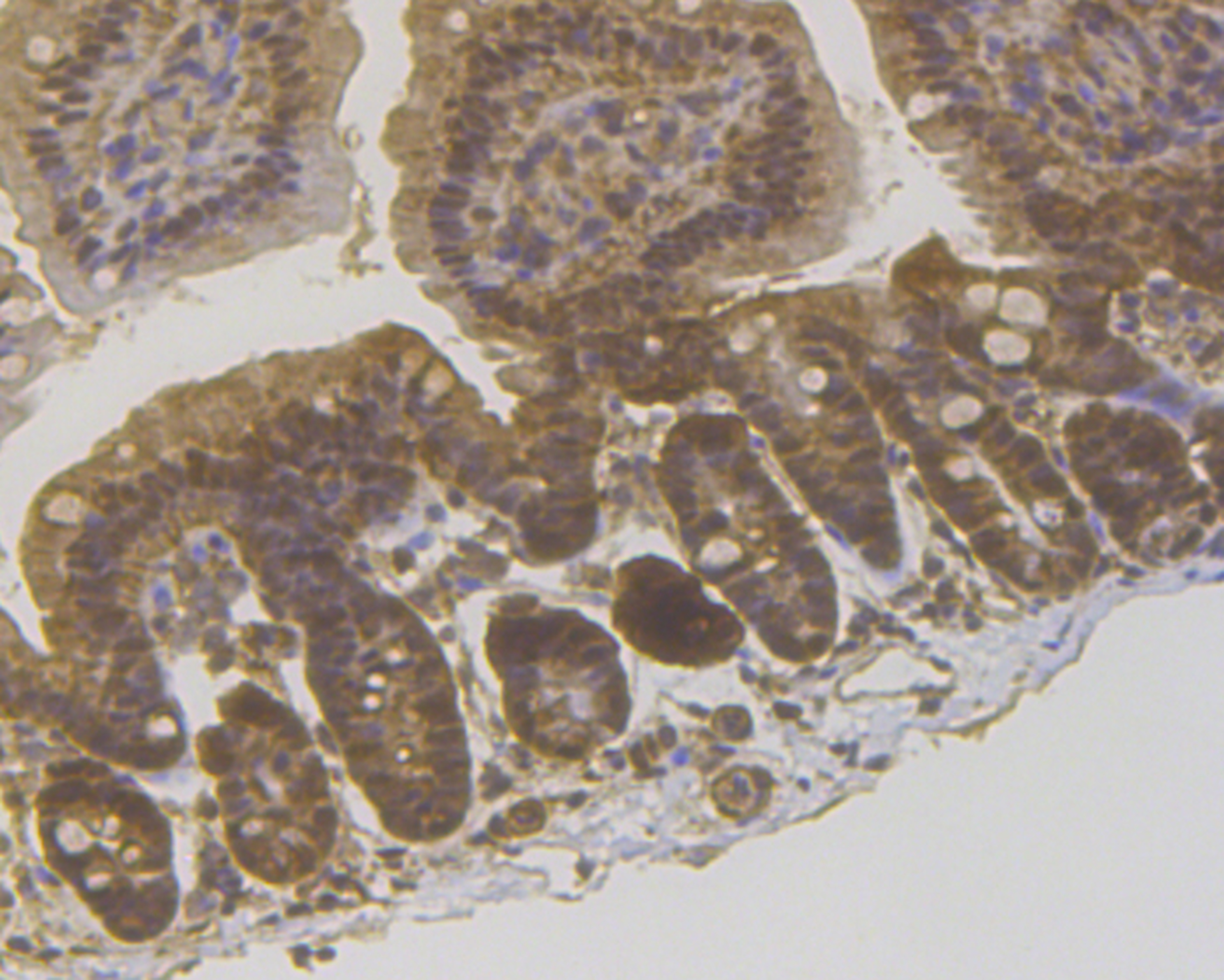
Fig5: Immunohistochemical analysis of paraffin-embedded mouse colon tissue using anti-PPP1CB antibody. The section was pre-treated using heat mediated antigen retrieval with sodium citrate buffer (pH 6.0) for 20 mins.The tissues were blocked in 5% BSA for 30 minutes at room temperature, washed with ddH2O and PBS, and then probed with the antibody (ET7109-40) at 1/200 dilution, for 30 minutes at room temperature and detected using an HRP conjugated compact polymer system. DAB was used as the chrogen. Counter stained with hematoxylin and mounted with DPX.Application
Fig5: Immunohistochemical analysis of paraffin-embedded mouse colon tissue using anti-PPP1CB antibody. The section was pre-treated using heat mediated antigen retrieval with sodium citrate buffer (pH 6.0) for 20 mins.The tissues were blocked in 5% BSA for 30 minutes at room temperature, washed with ddH2O and PBS, and then probed with the antibody (ET7109-40) at 1/200 dilution, for 30 minutes at room temperature and detected using an HRP conjugated compact polymer system. DAB was used as the chrogen. Counter stained with hematoxylin and mounted with DPX.Application
Fig5: Immunohistochemical analysis of paraffin-embedded mouse colon tissue using anti-PPP1CB antibody. The section was pre-treated using heat mediated antigen retrieval with sodium citrate buffer (pH 6.0) for 20 mins.The tissues were blocked in 5% BSA for 30 minutes at room temperature, washed with ddH2O and PBS, and then probed with the antibody (ET7109-40) at 1/200 dilution, for 30 minutes at room temperature and detected using an HRP conjugated compact polymer system. DAB was used as the chrogen. Counter stained with hematoxylin and mounted with DPX.Application
Fig5: Immunohistochemical analysis of paraffin-embedded mouse colon tissue using anti-PPP1CB antibody. The section was pre-treated using heat mediated antigen retrieval with sodium citrate buffer (pH 6.0) for 20 mins.The tissues were blocked in 5% BSA for 30 minutes at room temperature, washed with ddH2O and PBS, and then probed with the antibody (ET7109-40) at 1/200 dilution, for 30 minutes at room temperature and detected using an HRP conjugated compact polymer system. DAB was used as the chrogen. Counter stained with hematoxylin and mounted with DPX.Application
Fig5: Immunohistochemical analysis of paraffin-embedded mouse colon tissue using anti-PPP1CB antibody. The section was pre-treated using heat mediated antigen retrieval with sodium citrate buffer (pH 6.0) for 20 mins.The tissues were blocked in 5% BSA for 30 minutes at room temperature, washed with ddH2O and PBS, and then probed with the antibody (ET7109-40) at 1/200 dilution, for 30 minutes at room temperature and detected using an HRP conjugated compact polymer system. DAB was used as the chrogen. Counter stained with hematoxylin and mounted with DPX.| Product Name | PPP1CB [JE45-08] |
|---|---|
| Antibody Type | Primary Antibodies |
| Antigen Alias | MGC3672 antibody PP 1B antibody PP-1B antibody PP1B antibody PP1B_HUMAN antibody PP1beta antibody PPP1CB antibody PPP1CD antibody Protein phosphatase 1 beta antibody Protein phosphatase 1 catalytic subunit beta isoform antibody Protein phosphatase 1 delta antibody Protein phosphatase 1, catalytic subunit, beta isozyme antibody Protein phosphatase 1, catalytic subunit, delta isoform antibody Serine threonine protein phosphatase PP1 beta catalytic subunit antibody Serine/threonine-protein phosphatase PP1-beta catalytic subunit antibody |
| Product description | In eukaryotes, the phosphorylation and dephosphorylation of proteins on serine and threonine residues is an essential means of regulating a broad range of cellular functions, including division, homeostasis and apoptosis. A group of proteins that are intimately involved in this process are the protein phosphatases. In general, the protein phosphatase (PP) holoenzyme is a trimeric complex composed of a regulatory subunit, a variable subunit and a catalytic subunit. Four major families of protein phosphatase catalytic subunit have been identified, designated PP1, PP2A, PP2B (calcineurin) and PP2C. An additional protein phosphatase catalytic subunit, PPX (also known as PP4) is a putative member of a novel PP family. The PP1 family is comprised of subfamily members PP1α, PP1β and PP1γ, which are MgATP-dependent enzymes. PP1 inactivity is maintained through its association with the inhibitory protein NIPP-1 (nuclear inhibitor of PP1). Phosphorylation of NIPP-1 by cAMP-PK or casein kinase II results in the release of active PP1. |
| Immunogen | Recombinant protein corresponding to human PPP1CB aa 100-327. |
| Clonality | Monoclonal |
|---|---|
| Isotype | IgG |
| Host Species | Recombinant rabbit |
| Tested Applications | WBIPIHC |
| WB:1:1,000-1:10,000 IP:1:50 IHC:1:50-1:200 |
|
| Species Reactivity | HumanMouseRat |
| Concentration | 1mg/ml |
| Alternative Names | MGC3672 antibody
PP 1B antibody
PP-1B antibody
PP1B antibody
PP1B_HUMAN antibody
PP1beta antibody
PPP1CB antibody
PPP1CD antibody
Protein phosphatase 1 beta antibody
Protein phosphatase 1 catalytic subunit beta isoform antibody
Protein phosphatase 1 delta antibody
Protein phosphatase 1 catalytic subunit beta isozyme antibody Protein phosphatase 1 catalytic subunit delta isoform antibody Serine threonine protein phosphatase PP1 beta catalytic subunit antibody Serine/threonine-protein phosphatase PP1-beta catalytic subunit antibody |
|---|---|
| Molecular Weight(MW) | 37 kDa |
| Cellular Localization | Cytoplasm. Nucleus. |
Application
Fig5: Immunohistochemical analysis of paraffin-embedded mouse colon tissue using anti-PPP1CB antibody. The section was pre-treated using heat mediated antigen retrieval with sodium citrate buffer (pH 6.0) for 20 mins.The tissues were blocked in 5% BSA for 30 minutes at room temperature, washed with ddH2O and PBS, and then probed with the antibody (ET7109-40) at 1/200 dilution, for 30 minutes at room temperature and detected using an HRP conjugated compact polymer system. DAB was used as the chrogen. Counter stained with hematoxylin and mounted with DPX.
Application
Fig5: Immunohistochemical analysis of paraffin-embedded mouse colon tissue using anti-PPP1CB antibody. The section was pre-treated using heat mediated antigen retrieval with sodium citrate buffer (pH 6.0) for 20 mins.The tissues were blocked in 5% BSA for 30 minutes at room temperature, washed with ddH2O and PBS, and then probed with the antibody (ET7109-40) at 1/200 dilution, for 30 minutes at room temperature and detected using an HRP conjugated compact polymer system. DAB was used as the chrogen. Counter stained with hematoxylin and mounted with DPX.
Application
Fig5: Immunohistochemical analysis of paraffin-embedded mouse colon tissue using anti-PPP1CB antibody. The section was pre-treated using heat mediated antigen retrieval with sodium citrate buffer (pH 6.0) for 20 mins.The tissues were blocked in 5% BSA for 30 minutes at room temperature, washed with ddH2O and PBS, and then probed with the antibody (ET7109-40) at 1/200 dilution, for 30 minutes at room temperature and detected using an HRP conjugated compact polymer system. DAB was used as the chrogen. Counter stained with hematoxylin and mounted with DPX.
Application
Fig5: Immunohistochemical analysis of paraffin-embedded mouse colon tissue using anti-PPP1CB antibody. The section was pre-treated using heat mediated antigen retrieval with sodium citrate buffer (pH 6.0) for 20 mins.The tissues were blocked in 5% BSA for 30 minutes at room temperature, washed with ddH2O and PBS, and then probed with the antibody (ET7109-40) at 1/200 dilution, for 30 minutes at room temperature and detected using an HRP conjugated compact polymer system. DAB was used as the chrogen. Counter stained with hematoxylin and mounted with DPX.
Application
Fig5: Immunohistochemical analysis of paraffin-embedded mouse colon tissue using anti-PPP1CB antibody. The section was pre-treated using heat mediated antigen retrieval with sodium citrate buffer (pH 6.0) for 20 mins.The tissues were blocked in 5% BSA for 30 minutes at room temperature, washed with ddH2O and PBS, and then probed with the antibody (ET7109-40) at 1/200 dilution, for 30 minutes at room temperature and detected using an HRP conjugated compact polymer system. DAB was used as the chrogen. Counter stained with hematoxylin and mounted with DPX.| Positive Control | Mouse skeletal muscle tissue, rat brain tissue, A431, SH-SY-5Y, SiHa, 293, Hela, PC-3M, HepG2, A549, human appendix tissue, human esophagus tissue, mouse colon tissue. |
|---|---|
| Application Notes | WB:1:1,000-1:10,000 IP:1:50 IHC:1:50-1:200 |
| Form | Liquid |
|---|---|
| Storage Instructions | Store at +4℃ after thawing. Aliquot store at -20℃. Avoid repeated freeze / thaw cycles. |
| Storage Buffer | 1*TBS (pH7.4), 1%BSA, 50%Glycerol. Preservative: 0.05% Sodium Azide. |


