Application
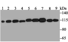
Fig1: Western blot analysis of RNF40 on different lysates. Proteins were transferred to a PVDF membrane and blocked with 5% BSA in PBS for 1 hour at room temperature. The primary antibody was used at a 1:500 dilution in 5% BSA at room temperature for 2 hours. Goat Anti-Rabbit IgG - HRP Secondary Antibody (HA1001) at 1:5,000 dilution was used for 1 hour at room temperature. Positive control: Lane 1: Mouse testis tissue lysate Lane 2: Rat brain tissue lysate Lane 3: A431 cell lysate Lane 4: SH-SY-5Y cell lysate Lane 5: SiHa cell lysate Lane 6: 293 cell lysate Lane 7: Hela cell lysate Lane 8: HepG2 cell lysate Lane 9: A549 cell lysateApplication
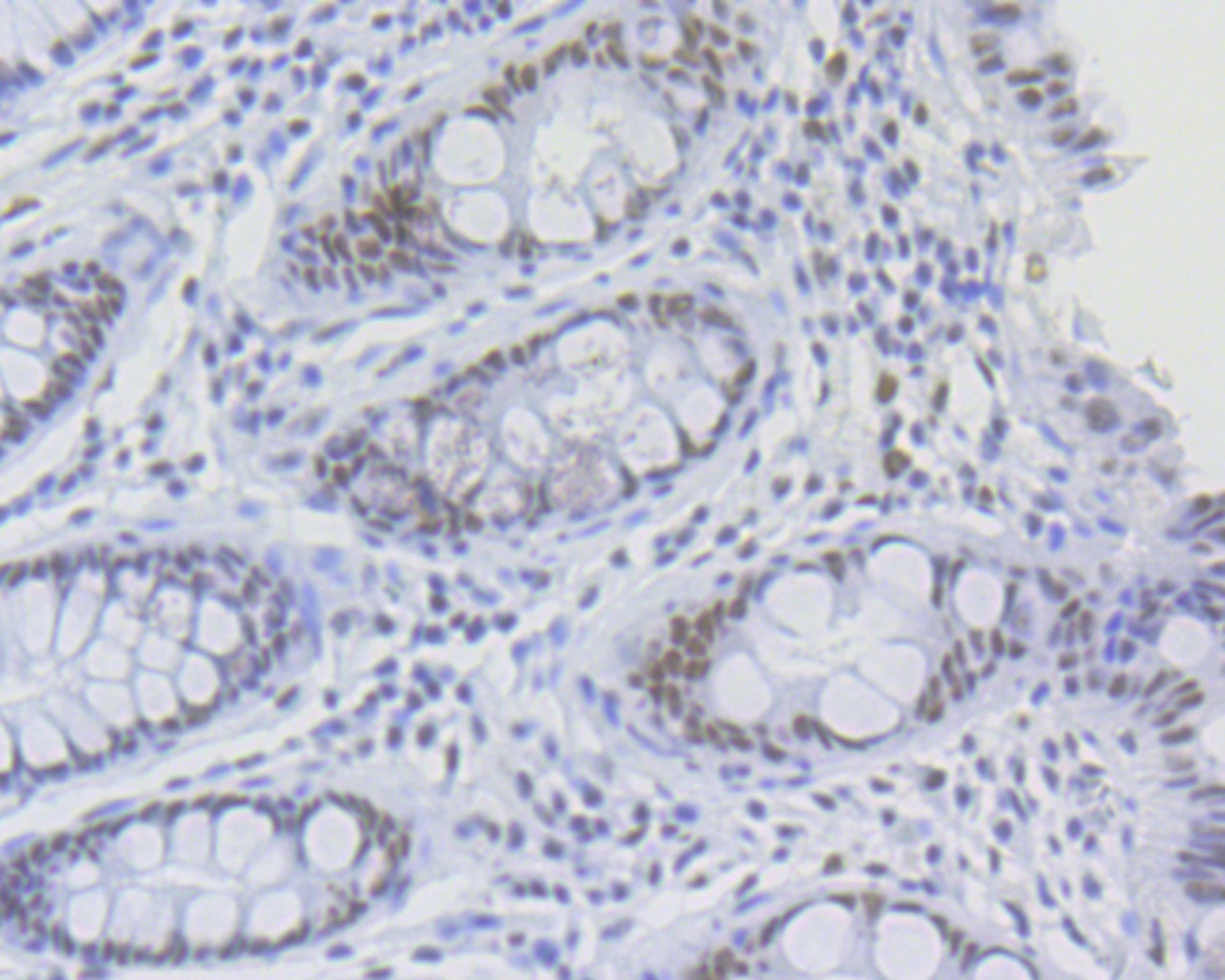
Fig2: Immunohistochemical analysis of paraffin-embedded human tonsil tissue using anti-RNF40 antibody. The section was pre-treated using heat mediated antigen retrieval with sodium citrate buffer (pH 6.0) for 20 mins. The tissues were blocked in 5% BSA for 30 minutes at room temperature, washed with ddH2O and PBS, and then probed with the antibody (ET7109-46) at 1/50 dilution, for 30 minutes at room temperature and detected using an HRP conjugated compact polymer system. DAB was used as the chrogen. Counter stained with hematoxylin and mounted with DPX.| Product Name | RNF40 [JE46-30] |
|---|---|
| Antibody Type | Primary Antibodies |
| Antigen Alias | 95 kDa retinoblastoma associated protein antibody 95 kDa retinoblastoma protein binding protein antibody 95 kDa retinoblastoma-associated protein antibody BRE 1B antibody BRE1 B antibody BRE1 E3 ubiquitin ligase homolog B antibody BRE1-B antibody BRE1B antibody BRE1B_HUMAN antibody DKFZp686K191 antibody E3 ubiquitin protein ligase BRE1B antibody E3 ubiquitin-protein ligase BRE1B antibody KIAA0661 antibody MGC13051 antibody Rb associated protein antibody RBP 95 antibody RBP95 antibody RING finger protein 40 antibody Ring finger protein 40 E3 ubiquitin protein ligase antibody RNF 40 antibody Rnf40 antibody STARING antibody |
| Product description | Component of the RNF20/40 E3 ubiquitin-protein ligase complex that mediates monoubiquitination of 'Lys-120' of histone H2B (H2BK120ub1). H2BK120ub1 gives a specific tag for epigenetic transcriptional activation and is also prerequisite for histone H3 'Lys-4' and 'Lys-79' methylation (H3K4me and H3K79me, respectively). It thereby plays a central role in histone code and gene regulation. The RNF20/40 complex forms a H2B ubiquitin ligase complex in cooperation with the E2 enzyme UBE2A or UBE2B; reports about the cooperation with UBE2E1/UBCH are contradictory. Required for transcriptional activation of Hox genes. This protein was reported to interact with the tumor suppressor protein RB1. Studies of the rat counterpart suggested that this protein may function as an E3 ubiquitin-protein ligase, and facilitate the ubiquitination and degradation of syntaxin 1, which is an essential component of the neurotransmitter release machinery. Multiple alternatively spliced transcript variants encoding different isoforms have been found for this gene. |
| Immunogen | Recombinant protein within human RNF40 aa 200-400. |
| Clonality | Monoclonal |
|---|---|
| Isotype | IgG |
| Host Species | Recombinant rabbit |
| Tested Applications | WBIPIHC |
| WB:1:1,000-1:2,000: IP:1:10-1:50: IHC:1:50: |
|
| Species Reactivity | HumanMouseRat |
| Concentration | 1mg/ml |
| Alternative Names | 95 kDa retinoblastoma associated protein antibody 95 kDa retinoblastoma protein binding protein antibody 95 kDa retinoblastoma-associated protein antibody BRE 1B antibody BRE1 B antibody BRE1 E3 ubiquitin ligase homolog B antibody BRE1-B antibody BRE1B antibody BRE1B_HUMAN antibody DKFZp686K191 antibody E3 ubiquitin protein ligase BRE1B antibody E3 ubiquitin-protein ligase BRE1B antibody KIAA0661 antibody MGC13051 antibody Rb associated protein antibody RBP 95 antibody RBP95 antibody RING finger protein 40 antibody Ring finger protein 40 E3 ubiquitin protein ligase antibody RNF 40 antibody Rnf40 antibody STARING antibody |
|---|---|
| Molecular Weight(MW) | 114 kDa |
| Cellular Localization | Nucleus. |
Application
Fig1: Western blot analysis of RNF40 on different lysates. Proteins were transferred to a PVDF membrane and blocked with 5% BSA in PBS for 1 hour at room temperature. The primary antibody was used at a 1:500 dilution in 5% BSA at room temperature for 2 hours. Goat Anti-Rabbit IgG - HRP Secondary Antibody (HA1001) at 1:5,000 dilution was used for 1 hour at room temperature. Positive control: Lane 1: Mouse testis tissue lysate Lane 2: Rat brain tissue lysate Lane 3: A431 cell lysate Lane 4: SH-SY-5Y cell lysate Lane 5: SiHa cell lysate Lane 6: 293 cell lysate Lane 7: Hela cell lysate Lane 8: HepG2 cell lysate Lane 9: A549 cell lysate
Application
Fig2: Immunohistochemical analysis of paraffin-embedded human tonsil tissue using anti-RNF40 antibody. The section was pre-treated using heat mediated antigen retrieval with sodium citrate buffer (pH 6.0) for 20 mins. The tissues were blocked in 5% BSA for 30 minutes at room temperature, washed with ddH2O and PBS, and then probed with the antibody (ET7109-46) at 1/50 dilution, for 30 minutes at room temperature and detected using an HRP conjugated compact polymer system. DAB was used as the chrogen. Counter stained with hematoxylin and mounted with DPX.| Positive Control | Mouse testis tissue, rat brain tissue, A431, SH-SY-5Y, SiHa, 293, Hela, HepG2, A549. |
|---|---|
| Application Notes | WB:1:1,000-1:2,000: IP:1:10-1:50: IHC:1:50: |
| Form | Liquid |
|---|---|
| Storage Instructions | Store at +4℃ after thawing. Aliquot store at -20℃. Avoid repeated freeze / thaw cycles. |
| Storage Buffer | 1*TBS (pH7.4), 1%BSA, 40%Glycerol. Preservative: 0.05% Sodium Azide. |


