Application
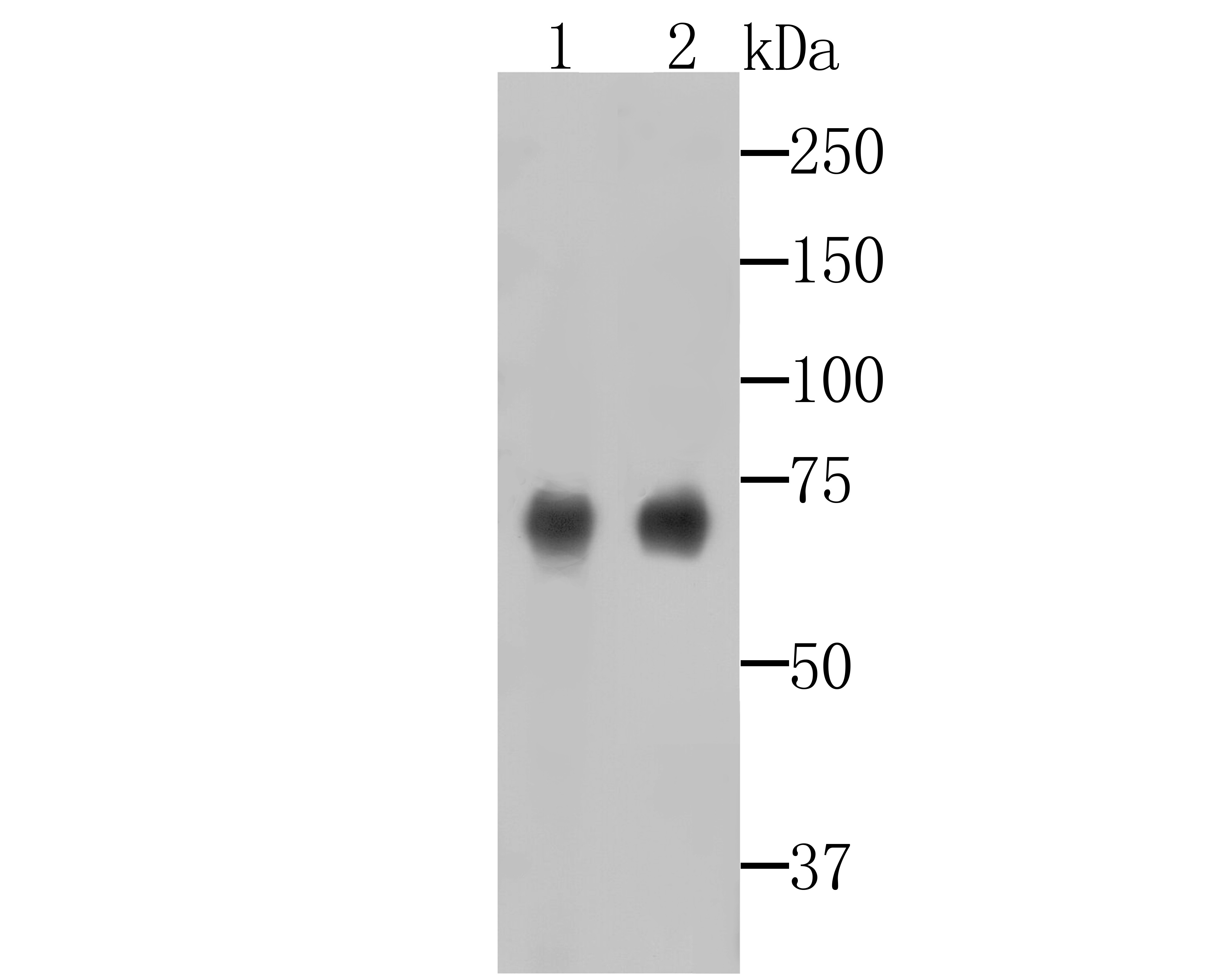
Fig1: Western blot analysis of SAM68 on different lysates. Proteins were transferred to a PVDF membrane and blocked with 5% BSA in PBS for 1 hour at room temperature. The primary antibody (ET7109-52, 1/1000) was used in 5% BSA at room temperature for 2 hours. Goat Anti-Rabbit IgG - HRP Secondary Antibody (HA1001) at 1:5,000 dilution was used for 1 hour at room temperature. Positive control: Lane 1: SHSY-5Y cell lysate Lane 2: Mouse brain tissue lysateApplication
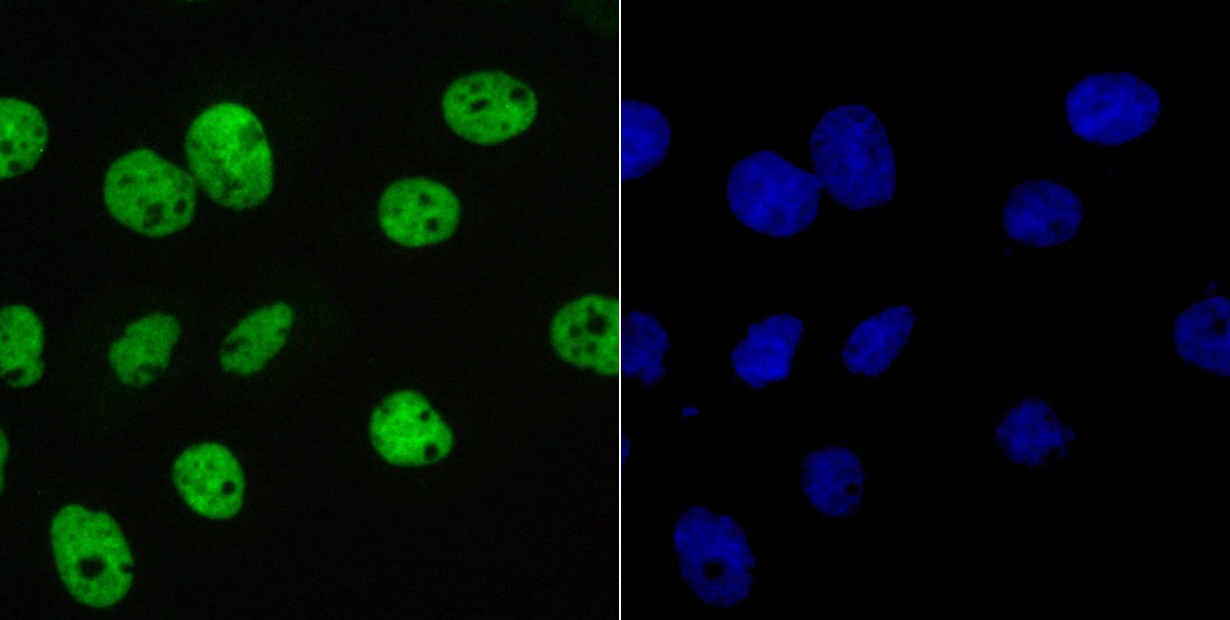
Fig2: ICC staining of SAM68 in A431 cells (green). Formalin fixed cells were permeabilized with 0.1% Triton X-100 in TBS for 10 minutes at room temperature and blocked with 1% Blocker BSA for 15 minutes at room temperature. Cells were probed with the primary antibody (ET7109-52, 1/50) for 1 hour at room temperature, washed with PBS. Alexa Fluor®488 Goat anti-Rabbit IgG was used as the secondary antibody at 1/100 dilution. The nuclear counter stain is DAPI (blue).Application
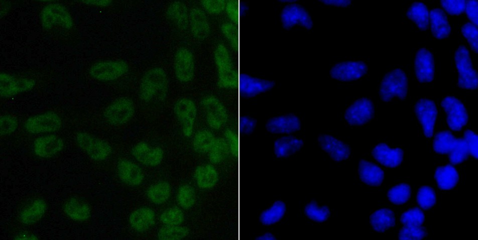
Fig3: ICC staining of SAM68 in F9 cells (green). Formalin fixed cells were permeabilized with 0.1% Triton X-100 in TBS for 10 minutes at room temperature and blocked with 1% Blocker BSA for 15 minutes at room temperature. Cells were probed with the primary antibody (ET7109-52, 1/50) for 1 hour at room temperature, washed with PBS. Alexa Fluor®488 Goat anti-Rabbit IgG was used as the secondary antibody at 1/100 dilution. The nuclear counter stain is DAPI (blue).Application
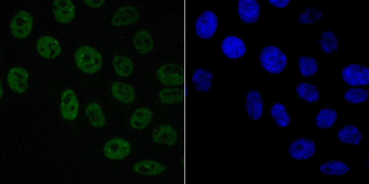
Fig4: ICC staining of SAM68 in HepG2 cells (green). Formalin fixed cells were permeabilized with 0.1% Triton X-100 in TBS for 10 minutes at room temperature and blocked with 1% Blocker BSA for 15 minutes at room temperature. Cells were probed with the primary antibody (ET7109-52, 1/50) for 1 hour at room temperature, washed with PBS. Alexa Fluor®488 Goat anti-Rabbit IgG was used as the secondary antibody at 1/100 dilution. The nuclear counter stain is DAPI (blue).Application
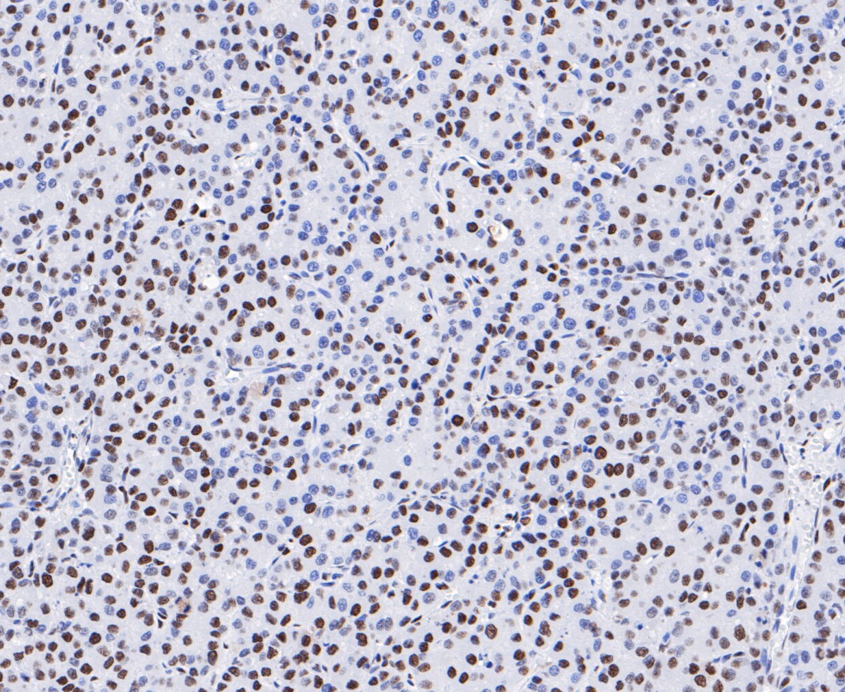
Fig5: Immunohistochemical analysis of paraffin-embedded human liver cancer tissue using anti-SAM68 antibody. The section was pre-treated using heat mediated antigen retrieval with sodium citrate buffer (pH 6.0) for 20 minutes. The tissues were blocked in 5% BSA for 30 minutes at room temperature, washed with ddH2O and PBS, and then probed with the primary antibody (ET7109-52, 1/1,000) for 30 minutes at room temperature. The detection was performed using an HRP conjugated compact polymer system. DAB was used as the chromogen. Tissues were counterstained with hematoxylin and mounted with DPX.Application
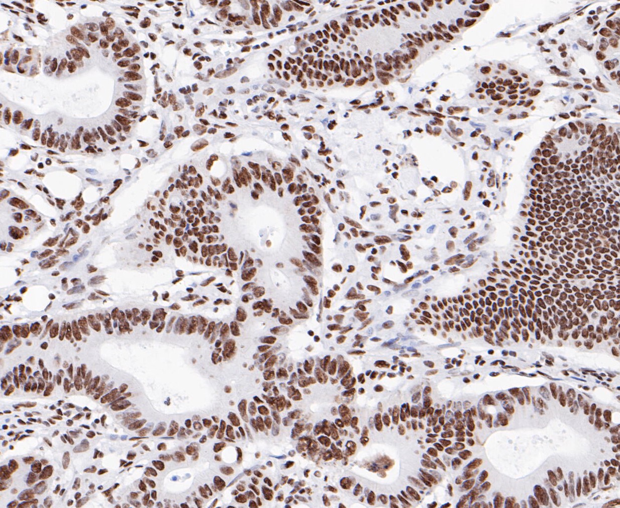
Fig6: Immunohistochemical analysis of paraffin-embedded human colon cancer tissue using anti-SAM68 antibody. The section was pre-treated using heat mediated antigen retrieval with sodium citrate buffer (pH 6.0) for 20 minutes. The tissues were blocked in 5% BSA for 30 minutes at room temperature, washed with ddH2O and PBS, and then probed with the primary antibody (ET7109-52, 1/1,000) for 30 minutes at room temperature. The detection was performed using an HRP conjugated compact polymer system. DAB was used as the chromogen. Tissues were counterstained with hematoxylin and mounted with DPX.Application
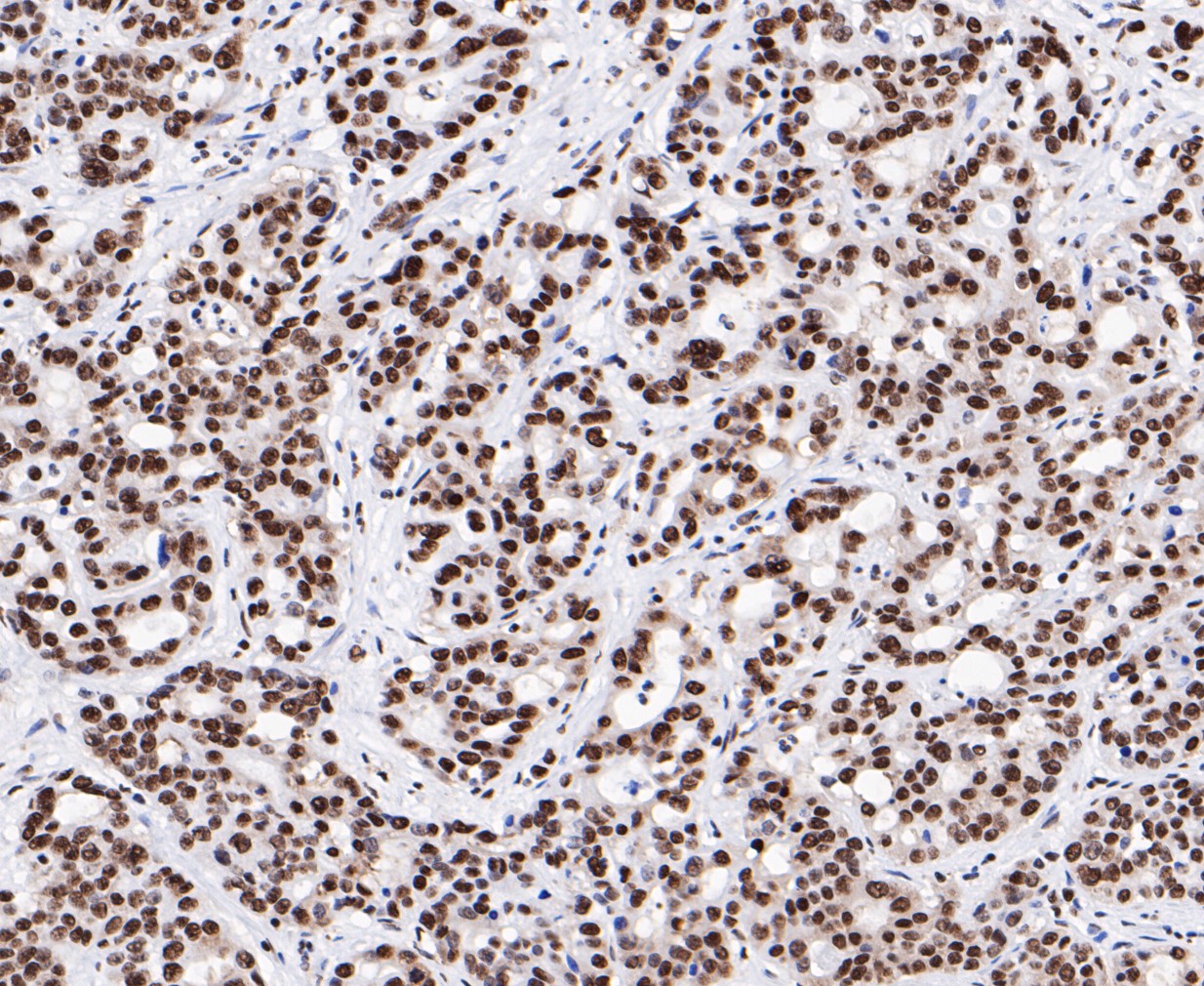
Fig7: Immunohistochemical analysis of paraffin-embedded human stomach cancer tissue using anti-SAM68 antibody. The section was pre-treated using heat mediated antigen retrieval with sodium citrate buffer (pH 6.0) for 20 minutes. The tissues were blocked in 5% BSA for 30 minutes at room temperature, washed with ddH2O and PBS, and then probed with the primary antibody (ET7109-52, 1/1,000) for 30 minutes at room temperature. The detection was performed using an HRP conjugated compact polymer system. DAB was used as the chromogen. Tissues were counterstained with hematoxylin and mounted with DPX.| Product Name | SAM68 [JE47-15] |
|---|---|
| Antibody Type | Primary Antibodies |
| Antigen Alias | FLJ34027 antibody GAP associated tyrosine phosphoprotein p62 antibody GAP-associated tyrosine phosphoprotein p62 antibody KH domain containing RNA binding signal transduction associated 1 antibody KH domain-containing antibody KHDR1_HUMAN antibody Khdrbs1 antibody p21 Ras GTPase activating protein associated p62 antibody p21 Ras GTPase-activating protein-associated p62 antibody p62 antibody p68 antibody RNA-binding antibody Sam68 antibody signal transduction-associated protein 1 antibody Src associated in mitosis 68 kDa protein antibody Src-associated in mitosis 68 kDa protein antibody |
| Product description | Recruited and tyrosine phosphorylated by several receptor systems, for example the T-cell, leptin and insulin receptors. Once phosphorylated, functions as an adapter protein in signal transduction cascades by binding to SH2 and SH3 domain-containing proteins. Role in G2-M progression in the cell cycle. Represses CBP-dependent transcriptional activation apparently by competing with other nuclear factors for binding to CBP. Also acts as a putative regulator of mRNA stability and/or translation rates and mediates mRNA nuclear export. Positively regulates the association of constitutive transport element (CTE)-containing mRNA with large polyribosomes and translation initiation. According to some authors, is not involved in the nucleocytoplasmic export of unspliced (CTE)-containing RNA species. RNA-binding protein that plays a role in the regulation of alternative splicing and influences mRNA splice site selection and exon inclusion. Binds to RNA containing 5'-[AU]UAA-3' as a bipartite motif spaced by more than 15 nucleotides. Binds poly(A). Can regulate CD44 alternative splicing in a Ras pathway-dependent manner. In cooperation with HNRNPA1 modulates alternative splicing of BCL2L1 by promoting splicing toward isoform Bcl-X(S), and of SMN1. Can regulate alternative splicing of NRXN1 and NRXN3 in the laminin G-like domain 6 containing the evolutionary conserved neurexin alternative spliced segment 4 (AS4) involved in neurexin selective targeting to postsynaptic partners. |
| Immunogen | Synthetic peptide within C terminal human SAM68. |
| Clonality | Monoclonal |
|---|---|
| Isotype | IgG |
| Host Species | Recombinant rabbit |
| Tested Applications | WBICCIHC |
| WB:1:1,000-1:5,000: ICC:1:50: IHC:1:1,000: |
|
| Species Reactivity | HumanMouseRat |
| Concentration | 1mg/ml |
| Alternative Names | FLJ34027 antibody GAP associated tyrosine phosphoprotein p62 antibody GAP-associated tyrosine phosphoprotein p62 antibody KH domain containing RNA binding signal transduction associated 1 antibody KH domain-containing antibody KHDR1_HUMAN antibody Khdrbs1 antibody p21 Ras GTPase activating protein associated p62 antibody p21 Ras GTPase-activating protein-associated p62 antibody p62 antibody p68 antibody RNA-binding antibody Sam68 antibody signal transduction-associated protein 1 antibody Src associated in mitosis 68 kDa protein antibody Src-associated in mitosis 68 kDa protein antibody |
|---|---|
| Molecular Weight(MW) | 68 kDa |
| Cellular Localization | Cytoplasm. Membrane. Nucleus. |
Application
Fig1: Western blot analysis of SAM68 on different lysates. Proteins were transferred to a PVDF membrane and blocked with 5% BSA in PBS for 1 hour at room temperature. The primary antibody (ET7109-52, 1/1000) was used in 5% BSA at room temperature for 2 hours. Goat Anti-Rabbit IgG - HRP Secondary Antibody (HA1001) at 1:5,000 dilution was used for 1 hour at room temperature. Positive control: Lane 1: SHSY-5Y cell lysate Lane 2: Mouse brain tissue lysate
Application
Fig2: ICC staining of SAM68 in A431 cells (green). Formalin fixed cells were permeabilized with 0.1% Triton X-100 in TBS for 10 minutes at room temperature and blocked with 1% Blocker BSA for 15 minutes at room temperature. Cells were probed with the primary antibody (ET7109-52, 1/50) for 1 hour at room temperature, washed with PBS. Alexa Fluor®488 Goat anti-Rabbit IgG was used as the secondary antibody at 1/100 dilution. The nuclear counter stain is DAPI (blue).
Application
Fig3: ICC staining of SAM68 in F9 cells (green). Formalin fixed cells were permeabilized with 0.1% Triton X-100 in TBS for 10 minutes at room temperature and blocked with 1% Blocker BSA for 15 minutes at room temperature. Cells were probed with the primary antibody (ET7109-52, 1/50) for 1 hour at room temperature, washed with PBS. Alexa Fluor®488 Goat anti-Rabbit IgG was used as the secondary antibody at 1/100 dilution. The nuclear counter stain is DAPI (blue).
Application
Fig4: ICC staining of SAM68 in HepG2 cells (green). Formalin fixed cells were permeabilized with 0.1% Triton X-100 in TBS for 10 minutes at room temperature and blocked with 1% Blocker BSA for 15 minutes at room temperature. Cells were probed with the primary antibody (ET7109-52, 1/50) for 1 hour at room temperature, washed with PBS. Alexa Fluor®488 Goat anti-Rabbit IgG was used as the secondary antibody at 1/100 dilution. The nuclear counter stain is DAPI (blue).
Application
Fig5: Immunohistochemical analysis of paraffin-embedded human liver cancer tissue using anti-SAM68 antibody. The section was pre-treated using heat mediated antigen retrieval with sodium citrate buffer (pH 6.0) for 20 minutes. The tissues were blocked in 5% BSA for 30 minutes at room temperature, washed with ddH2O and PBS, and then probed with the primary antibody (ET7109-52, 1/1,000) for 30 minutes at room temperature. The detection was performed using an HRP conjugated compact polymer system. DAB was used as the chromogen. Tissues were counterstained with hematoxylin and mounted with DPX.
Application
Fig6: Immunohistochemical analysis of paraffin-embedded human colon cancer tissue using anti-SAM68 antibody. The section was pre-treated using heat mediated antigen retrieval with sodium citrate buffer (pH 6.0) for 20 minutes. The tissues were blocked in 5% BSA for 30 minutes at room temperature, washed with ddH2O and PBS, and then probed with the primary antibody (ET7109-52, 1/1,000) for 30 minutes at room temperature. The detection was performed using an HRP conjugated compact polymer system. DAB was used as the chromogen. Tissues were counterstained with hematoxylin and mounted with DPX.
Application
Fig7: Immunohistochemical analysis of paraffin-embedded human stomach cancer tissue using anti-SAM68 antibody. The section was pre-treated using heat mediated antigen retrieval with sodium citrate buffer (pH 6.0) for 20 minutes. The tissues were blocked in 5% BSA for 30 minutes at room temperature, washed with ddH2O and PBS, and then probed with the primary antibody (ET7109-52, 1/1,000) for 30 minutes at room temperature. The detection was performed using an HRP conjugated compact polymer system. DAB was used as the chromogen. Tissues were counterstained with hematoxylin and mounted with DPX.| Positive Control | SHSY-5Y, mouse brain tissue, A431, F9, HepG2, human liver cancer tissue, human colon cancer tissue, human stomach cancer tissue. |
|---|---|
| Application Notes | WB:1:1,000-1:5,000: ICC:1:50: IHC:1:1,000: |
| Form | Liquid |
|---|---|
| Storage Instructions | Store at +4℃ after thawing. Aliquot store at -20℃. Avoid repeated freeze / thaw cycles. |
| Storage Buffer | 1*TBS (pH7.4), 1%BSA, 50%Glycerol. Preservative: 0.05% Sodium Azide. |


