Application
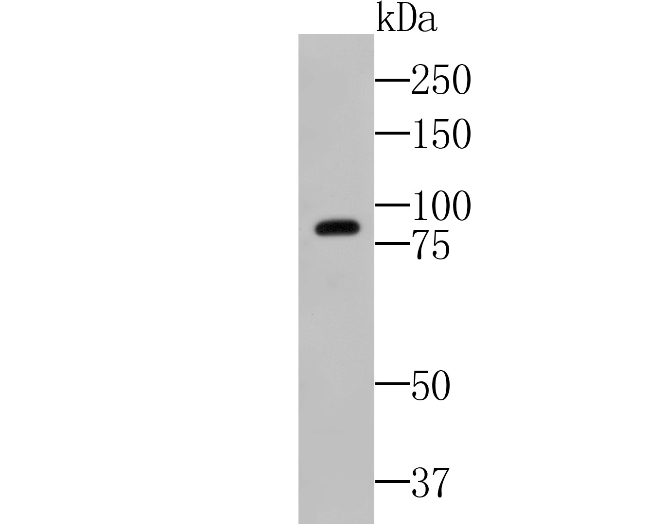
Fig1: Western blot analysis of GTPBP4 on 293T cell lysates. Proteins were transferred to a PVDF membrane and blocked with 5% BSA in PBS for 1 hour at room temperature. The primary antibody (ET7109-59, 1/500) was used in 5% BSA at room temperature for 2 hours. Goat Anti-Rabbit IgG - HRP Secondary Antibody (HA1001) at 1:5,000 dilution was used for 1 hour at room temperature.Application
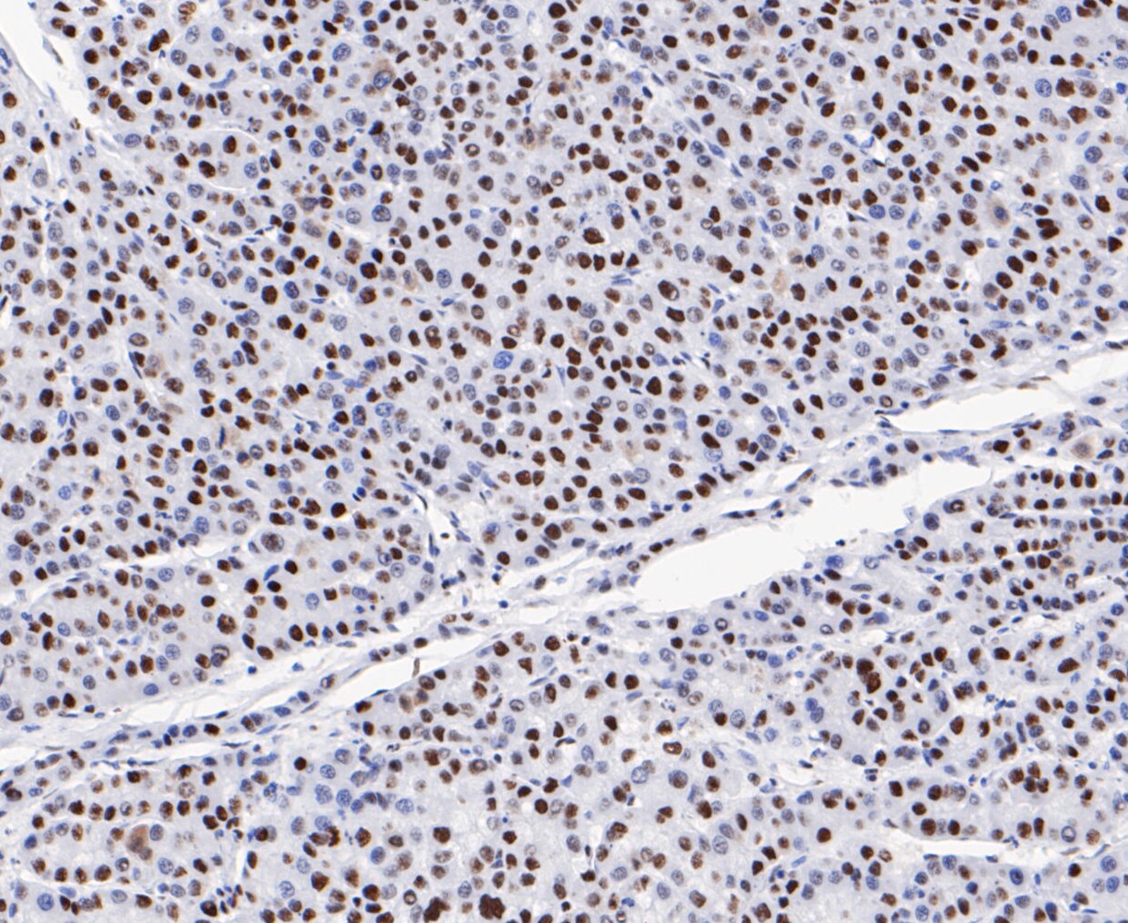
Fig2: Immunohistochemical analysis of paraffin-embedded human liver cancer tissue using anti-GTPBP4 antibody. The section was pre-treated using heat mediated antigen retrieval with sodium citrate buffer (pH 6.0) for 20 minutes. The tissues were blocked in 5% BSA for 30 minutes at room temperature, washed with ddH2O and PBS, and then probed with the primary antibody (ET7109-59, 1/50) for 30 minutes at room temperature. The detection was performed using an HRP conjugated compact polymer system. DAB was used as the chromogen. Tissues were counterstained with hematoxylin and mounted with DPX.Application
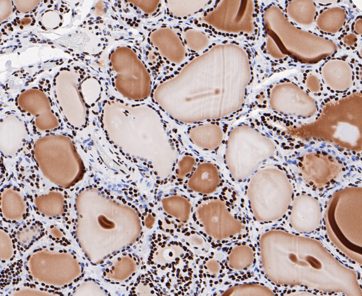
Fig3: Immunohistochemical analysis of paraffin-embedded human thyroid gland tissue using anti-GTPBP4 antibody. The section was pre-treated using heat mediated antigen retrieval with sodium citrate buffer (pH 6.0) for 20 minutes. The tissues were blocked in 5% BSA for 30 minutes at room temperature, washed with ddH2O and PBS, and then probed with the primary antibody (ET7109-59, 1/50) for 30 minutes at room temperature. The detection was performed using an HRP conjugated compact polymer system. DAB was used as the chromogen. Tissues were counterstained with hematoxylin and mounted with DPX.Application
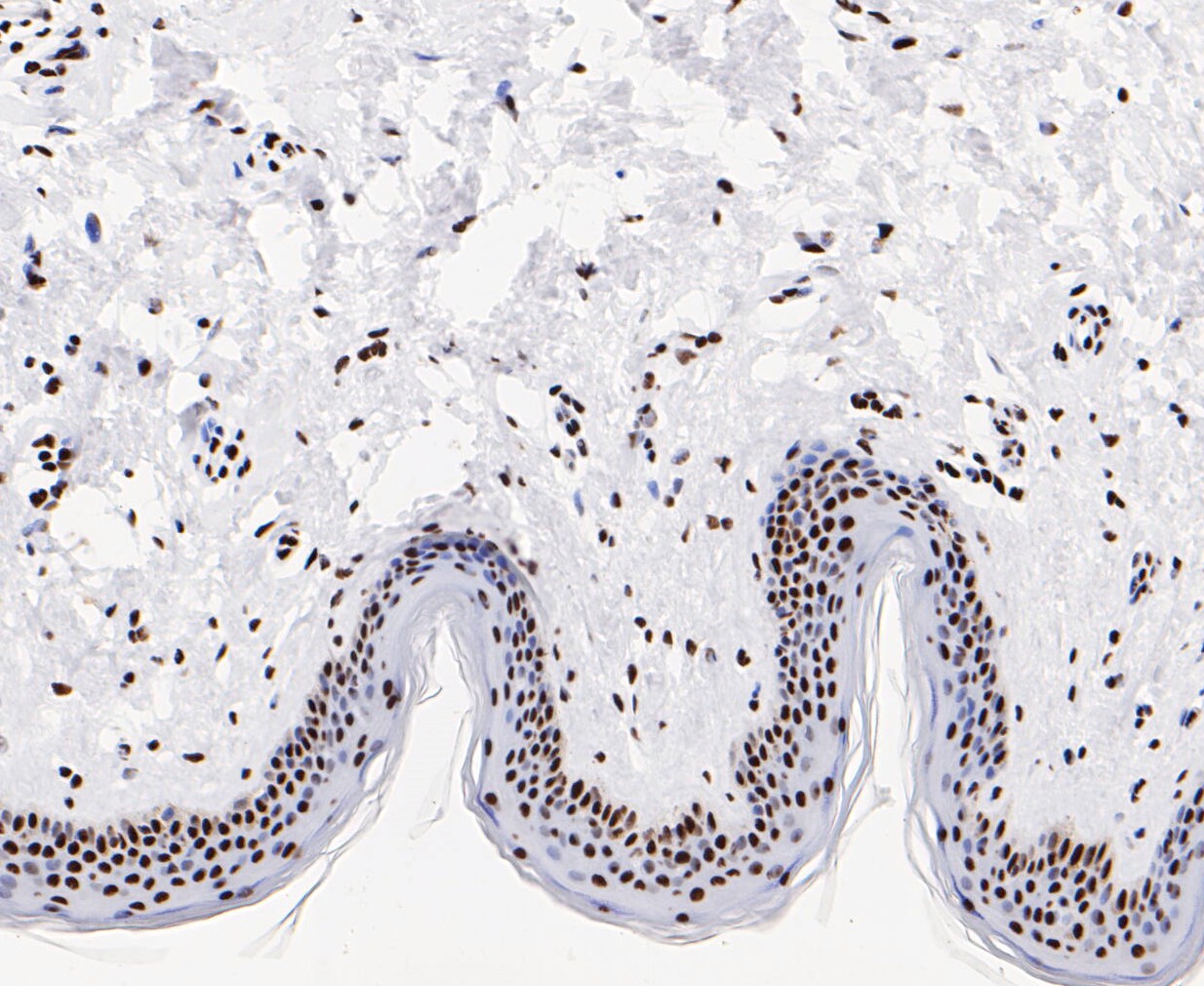
Fig4: Immunohistochemical analysis of paraffin-embedded human skin tissue using anti-GTPBP4 antibody. The section was pre-treated using heat mediated antigen retrieval with sodium citrate buffer (pH 6.0) for 20 minutes. The tissues were blocked in 5% BSA for 30 minutes at room temperature, washed with ddH2O and PBS, and then probed with the primary antibody (ET7109-59, 1/50) for 30 minutes at room temperature. The detection was performed using an HRP conjugated compact polymer system. DAB was used as the chromogen. Tissues were counterstained with hematoxylin and mounted with DPX.Application
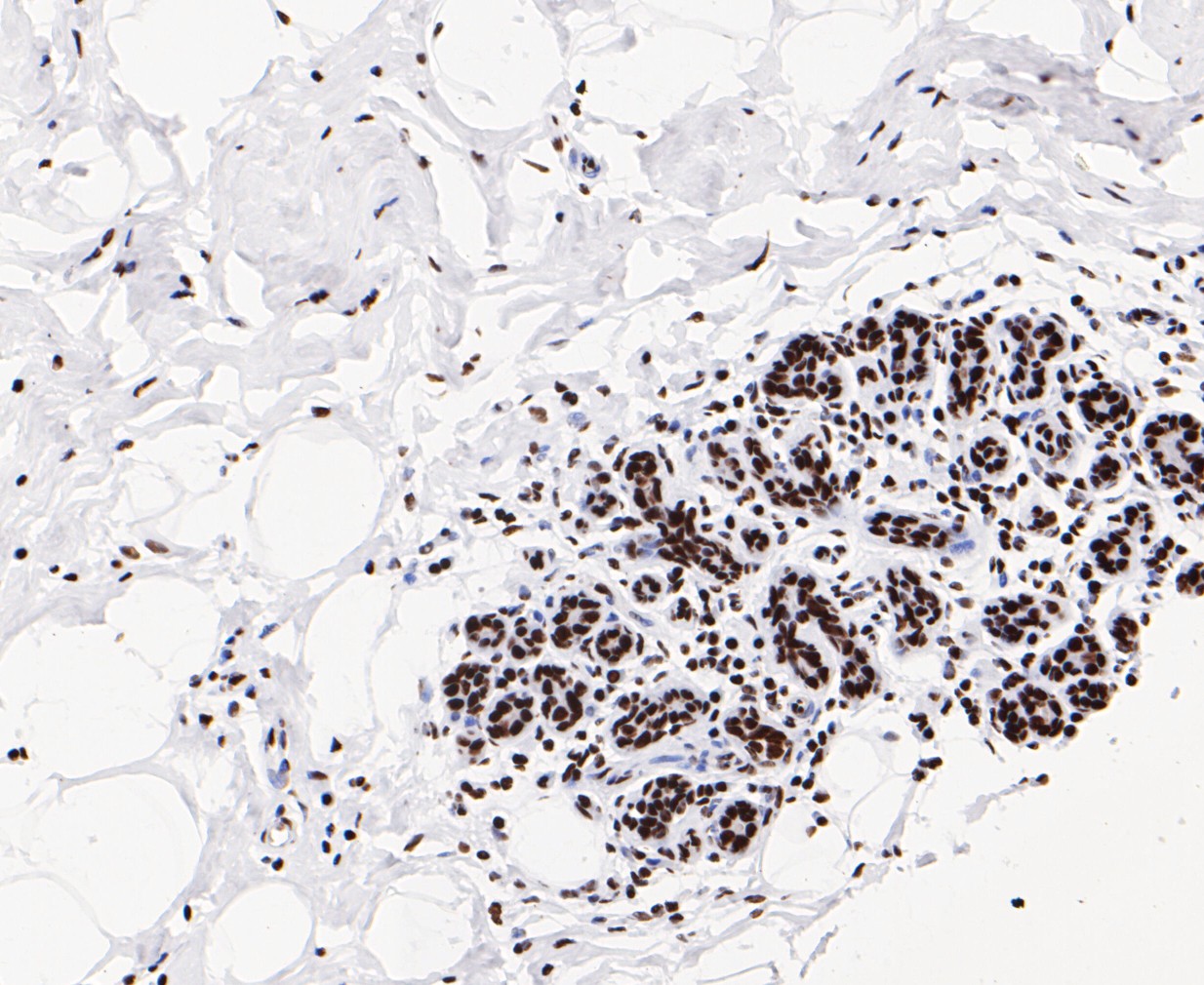
Fig5: Immunohistochemical analysis of paraffin-embedded human breast tissue using anti-GTPBP4 antibody. The section was pre-treated using heat mediated antigen retrieval with sodium citrate buffer (pH 6.0) for 20 minutes. The tissues were blocked in 5% BSA for 30 minutes at room temperature, washed with ddH2O and PBS, and then probed with the primary antibody (ET7109-59, 1/50) for 30 minutes at room temperature. The detection was performed using an HRP conjugated compact polymer system. DAB was used as the chromogen. Tissues were counterstained with hematoxylin and mounted with DPX.Application
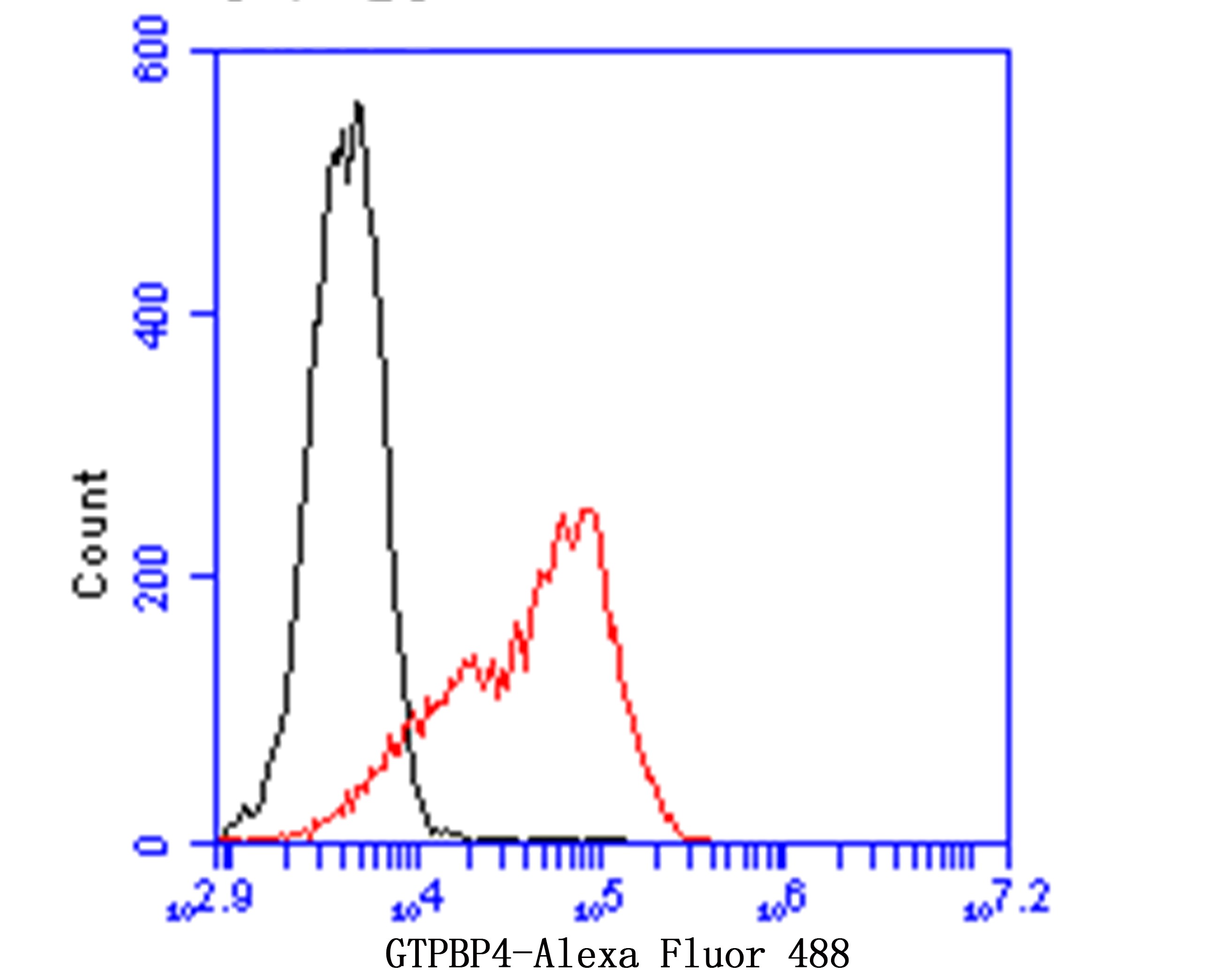
Fig6: Flow cytometric analysis of GTPBP4 was done on thp-1 cells. The cells were fixed, permeabilized and stained with the primary antibody (ET7109-59, 1/100) (red). After incubation of the primary antibody at room temperature for an hour, the cells were stained with a Alexa Fluor 488-conjugated goat anti-rabbit IgG Secondary antibody at 1/500 dilution for 30 minutes.Unlabelled sample was used as a control (cells without incubation with primary antibody; black).| Product Name | GTPBP4 [JE47-47] |
|---|---|
| Antibody Type | Primary Antibodies |
| Antigen Alias | Chronic renal failure gene protein antibody CRFG antibody G protein binding protein CRFG antibody GTP binding protein 4 antibody GTP-binding protein NGB antibody Gtpbp4 antibody NGB antibody NOG1 antibody NOG1_HUMAN antibody Nucleolar GTP binding protein 1 antibody Nucleolar GTP-binding protein 1 antibody |
| Product description | GTP-binding proteins are GTPases and function as molecular switches that can flip between two states: active, when GTP is bound, and inactive, when GDP is bound. 'Active' in this context usually means that the molecule acts as a signal to trigger other events in the cell. When an extracellular ligand binds to a G-protein-linked receptor, the receptor changes its conformation and switches on the trimeric G proteins that associate with it by causing them to eject their GDP and replace it with GTP. The switch is turned off when the G protein hydrolyzes its own bound GTP, converting it back to GDP. But before that occurs, the active protein has an opportunity to diffuse away from the receptor and deliver its message for a prolonged period to its downstream target. |
| Immunogen | Synthetic peptide within N terminal human GTPBP4. |
| Clonality | Monoclonal |
|---|---|
| Isotype | IgG |
| Host Species | Recombinant rabbit |
| Tested Applications | WBIHCFC |
| WB:1:500: IHC:1:50-1:200: FC:1:50-1:100: |
|
| Species Reactivity | Human |
| Concentration | 1mg/ml |
| Alternative Names | Chronic renal failure gene protein antibody CRFG antibody G protein binding protein CRFG antibody GTP binding protein 4 antibody GTP-binding protein NGB antibody Gtpbp4 antibody NGB antibody NOG1 antibody NOG1_HUMAN antibody Nucleolar GTP binding protein 1 antibody Nucleolar GTP-binding protein 1 antibody |
|---|---|
| Molecular Weight(MW) | 74 kDa |
| Cellular Localization | Nucleus. |
| SwissProt ID | Q9BZE4 |
|---|
Application
Fig1: Western blot analysis of GTPBP4 on 293T cell lysates. Proteins were transferred to a PVDF membrane and blocked with 5% BSA in PBS for 1 hour at room temperature. The primary antibody (ET7109-59, 1/500) was used in 5% BSA at room temperature for 2 hours. Goat Anti-Rabbit IgG - HRP Secondary Antibody (HA1001) at 1:5,000 dilution was used for 1 hour at room temperature.
Application
Fig2: Immunohistochemical analysis of paraffin-embedded human liver cancer tissue using anti-GTPBP4 antibody. The section was pre-treated using heat mediated antigen retrieval with sodium citrate buffer (pH 6.0) for 20 minutes. The tissues were blocked in 5% BSA for 30 minutes at room temperature, washed with ddH2O and PBS, and then probed with the primary antibody (ET7109-59, 1/50) for 30 minutes at room temperature. The detection was performed using an HRP conjugated compact polymer system. DAB was used as the chromogen. Tissues were counterstained with hematoxylin and mounted with DPX.
Application
Fig3: Immunohistochemical analysis of paraffin-embedded human thyroid gland tissue using anti-GTPBP4 antibody. The section was pre-treated using heat mediated antigen retrieval with sodium citrate buffer (pH 6.0) for 20 minutes. The tissues were blocked in 5% BSA for 30 minutes at room temperature, washed with ddH2O and PBS, and then probed with the primary antibody (ET7109-59, 1/50) for 30 minutes at room temperature. The detection was performed using an HRP conjugated compact polymer system. DAB was used as the chromogen. Tissues were counterstained with hematoxylin and mounted with DPX.
Application
Fig4: Immunohistochemical analysis of paraffin-embedded human skin tissue using anti-GTPBP4 antibody. The section was pre-treated using heat mediated antigen retrieval with sodium citrate buffer (pH 6.0) for 20 minutes. The tissues were blocked in 5% BSA for 30 minutes at room temperature, washed with ddH2O and PBS, and then probed with the primary antibody (ET7109-59, 1/50) for 30 minutes at room temperature. The detection was performed using an HRP conjugated compact polymer system. DAB was used as the chromogen. Tissues were counterstained with hematoxylin and mounted with DPX.
Application
Fig5: Immunohistochemical analysis of paraffin-embedded human breast tissue using anti-GTPBP4 antibody. The section was pre-treated using heat mediated antigen retrieval with sodium citrate buffer (pH 6.0) for 20 minutes. The tissues were blocked in 5% BSA for 30 minutes at room temperature, washed with ddH2O and PBS, and then probed with the primary antibody (ET7109-59, 1/50) for 30 minutes at room temperature. The detection was performed using an HRP conjugated compact polymer system. DAB was used as the chromogen. Tissues were counterstained with hematoxylin and mounted with DPX.
Application
Fig6: Flow cytometric analysis of GTPBP4 was done on thp-1 cells. The cells were fixed, permeabilized and stained with the primary antibody (ET7109-59, 1/100) (red). After incubation of the primary antibody at room temperature for an hour, the cells were stained with a Alexa Fluor 488-conjugated goat anti-rabbit IgG Secondary antibody at 1/500 dilution for 30 minutes.Unlabelled sample was used as a control (cells without incubation with primary antibody; black).| Positive Control | 293T cell, human liver cancer tissue, human thyroid gland tissue, human skin tissue, human breast tissue, thp-1. |
|---|---|
| Application Notes | WB:1:500: IHC:1:50-1:200: FC:1:50-1:100: |
| Form | Liquid |
|---|---|
| Storage Instructions | Store at +4℃ after thawing. Aliquot store at -20℃. Avoid repeated freeze / thaw cycles. |
| Storage Buffer | 1*TBS (pH7.4), 1%BSA, 50%Glycerol. Preservative: 0.05% Sodium Azide. |


