Application
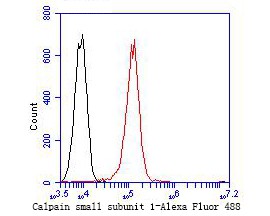
Fig7: Flow cytometric analysis of Calpain small subunit 1 was done on A549 cells. The cells were fixed, permeabilized and stained with the primary antibody (ET7109-66, 1/50) (red). After incubation of the primary antibody at room temperature for an hour, the cells were stained with a Alexa Fluor 488-conjugated Goat anti-Rabbit IgG Secondary antibody at 1/1000 dilution for 30 minutes.Unlabelled sample was used as a control (cells without incubation with primary antibody; black).Application
Fig7: Flow cytometric analysis of Calpain small subunit 1 was done on A549 cells. The cells were fixed, permeabilized and stained with the primary antibody (ET7109-66, 1/50) (red). After incubation of the primary antibody at room temperature for an hour, the cells were stained with a Alexa Fluor 488-conjugated Goat anti-Rabbit IgG Secondary antibody at 1/1000 dilution for 30 minutes.Unlabelled sample was used as a control (cells without incubation with primary antibody; black).Application
Fig7: Flow cytometric analysis of Calpain small subunit 1 was done on A549 cells. The cells were fixed, permeabilized and stained with the primary antibody (ET7109-66, 1/50) (red). After incubation of the primary antibody at room temperature for an hour, the cells were stained with a Alexa Fluor 488-conjugated Goat anti-Rabbit IgG Secondary antibody at 1/1000 dilution for 30 minutes.Unlabelled sample was used as a control (cells without incubation with primary antibody; black).Application
Fig7: Flow cytometric analysis of Calpain small subunit 1 was done on A549 cells. The cells were fixed, permeabilized and stained with the primary antibody (ET7109-66, 1/50) (red). After incubation of the primary antibody at room temperature for an hour, the cells were stained with a Alexa Fluor 488-conjugated Goat anti-Rabbit IgG Secondary antibody at 1/1000 dilution for 30 minutes.Unlabelled sample was used as a control (cells without incubation with primary antibody; black).Application
Fig7: Flow cytometric analysis of Calpain small subunit 1 was done on A549 cells. The cells were fixed, permeabilized and stained with the primary antibody (ET7109-66, 1/50) (red). After incubation of the primary antibody at room temperature for an hour, the cells were stained with a Alexa Fluor 488-conjugated Goat anti-Rabbit IgG Secondary antibody at 1/1000 dilution for 30 minutes.Unlabelled sample was used as a control (cells without incubation with primary antibody; black).Application
Fig7: Flow cytometric analysis of Calpain small subunit 1 was done on A549 cells. The cells were fixed, permeabilized and stained with the primary antibody (ET7109-66, 1/50) (red). After incubation of the primary antibody at room temperature for an hour, the cells were stained with a Alexa Fluor 488-conjugated Goat anti-Rabbit IgG Secondary antibody at 1/1000 dilution for 30 minutes.Unlabelled sample was used as a control (cells without incubation with primary antibody; black).Application
Fig7: Flow cytometric analysis of Calpain small subunit 1 was done on A549 cells. The cells were fixed, permeabilized and stained with the primary antibody (ET7109-66, 1/50) (red). After incubation of the primary antibody at room temperature for an hour, the cells were stained with a Alexa Fluor 488-conjugated Goat anti-Rabbit IgG Secondary antibody at 1/1000 dilution for 30 minutes.Unlabelled sample was used as a control (cells without incubation with primary antibody; black).| Product Name | Calpain small subunit 1 [JE47-72] |
|---|---|
| Antibody Type | Primary Antibodies |
| Antigen Alias | 30K antibody Calcium activated neutral proteinase antibody Calcium activated neutral proteinase small subunit antibody Calcium dependent protease small subunit 1 antibody Calcium dependent protease small subunit antibody Calcium-activated neutral proteinase small subunit antibody Calcium-dependent protease small subunit 1 antibody Calcium-dependent protease small subunit antibody Calpain 4 antibody Calpain 4 small subunit (30K) antibody Calpain 4 small subunit antibody Calpain regulatory subunit antibody Calpain S1 antibody Calpain small polypeptide antibody Calpain small subunit 1 antibody Calpain4 antibody CalpainS1 antibody CANP antibody CANP small subunit antibody CANPS antibody CAPN 4 antibody CAPN S1 antibody CAPN4 antibody CAPNS 1 antibody CAPNS antibody Capns1 antibody CDPS antibody CPNS1_HUMAN antibody CSS 1 antibody CSS1 antibody |
| Product description | This gene is a member of the calpain small subunit family. Calpains are calcium-dependent cysteine proteinases that are widely distributed in mammalian cells. Calpains operate as heterodimers, comprising a specific large catalytic subunit (calpain 1 subunit in Calpain I, and calpain 2 subunit in Calpain II), and a common small regulatory subunit encoded by this gene. This encoded protein is essential for the stability and function of both calpain heterodimers, whose proteolytic activities influence various cellular functions including apoptosis, proliferation, migration, adhesion, and autophagy. Calpains have been implicated in neurodegenerative processes, such as myotonic dystrophy. A pseudogene of this gene has been defined on chromosome 1. Alternative splicing results in multiple transcript variants. |
| Immunogen | Recombinant protein within N-terminal human Calpain small subunit 1. |
| Clonality | Monoclonal |
|---|---|
| Isotype | IgG |
| Host Species | Recombinant rabbit |
| Tested Applications | WBIHCFC |
| WB:1:500-1:2,000: IHC:1:50-1:200: FC:1:50-1:100: |
|
| Species Reactivity | HumanMouseRat |
| Concentration | 1mg/ml |
| Alternative Names | 30K antibody Calcium activated neutral proteinase antibody Calcium activated neutral proteinase small subunit antibody Calcium dependent protease small subunit 1 antibody Calcium dependent protease small subunit antibody Calcium-activated neutral proteinase small subunit antibody Calcium-dependent protease small subunit 1 antibody Calcium-dependent protease small subunit antibody Calpain 4 antibody Calpain 4 small subunit (30K) antibody Calpain 4 small subunit antibody Calpain regulatory subunit antibody Calpain S1 antibody Calpain small polypeptide antibody Calpain small subunit 1 antibody Calpain4 antibody CalpainS1 antibody CANP antibody CANP small subunit antibody CANPS antibody CAPN 4 antibody CAPN S1 antibody CAPN4 antibody CAPNS 1 antibody CAPNS antibody Capns1 antibody CDPS antibody CPNS1_HUMAN antibody CSS 1 antibody CSS1 antibody |
|---|---|
| Molecular Weight(MW) | 28 kDa |
| Cellular Localization | Cytoplasm. Membrane. |
Application
Fig7: Flow cytometric analysis of Calpain small subunit 1 was done on A549 cells. The cells were fixed, permeabilized and stained with the primary antibody (ET7109-66, 1/50) (red). After incubation of the primary antibody at room temperature for an hour, the cells were stained with a Alexa Fluor 488-conjugated Goat anti-Rabbit IgG Secondary antibody at 1/1000 dilution for 30 minutes.Unlabelled sample was used as a control (cells without incubation with primary antibody; black).
Application
Fig7: Flow cytometric analysis of Calpain small subunit 1 was done on A549 cells. The cells were fixed, permeabilized and stained with the primary antibody (ET7109-66, 1/50) (red). After incubation of the primary antibody at room temperature for an hour, the cells were stained with a Alexa Fluor 488-conjugated Goat anti-Rabbit IgG Secondary antibody at 1/1000 dilution for 30 minutes.Unlabelled sample was used as a control (cells without incubation with primary antibody; black).
Application
Fig7: Flow cytometric analysis of Calpain small subunit 1 was done on A549 cells. The cells were fixed, permeabilized and stained with the primary antibody (ET7109-66, 1/50) (red). After incubation of the primary antibody at room temperature for an hour, the cells were stained with a Alexa Fluor 488-conjugated Goat anti-Rabbit IgG Secondary antibody at 1/1000 dilution for 30 minutes.Unlabelled sample was used as a control (cells without incubation with primary antibody; black).
Application
Fig7: Flow cytometric analysis of Calpain small subunit 1 was done on A549 cells. The cells were fixed, permeabilized and stained with the primary antibody (ET7109-66, 1/50) (red). After incubation of the primary antibody at room temperature for an hour, the cells were stained with a Alexa Fluor 488-conjugated Goat anti-Rabbit IgG Secondary antibody at 1/1000 dilution for 30 minutes.Unlabelled sample was used as a control (cells without incubation with primary antibody; black).
Application
Fig7: Flow cytometric analysis of Calpain small subunit 1 was done on A549 cells. The cells were fixed, permeabilized and stained with the primary antibody (ET7109-66, 1/50) (red). After incubation of the primary antibody at room temperature for an hour, the cells were stained with a Alexa Fluor 488-conjugated Goat anti-Rabbit IgG Secondary antibody at 1/1000 dilution for 30 minutes.Unlabelled sample was used as a control (cells without incubation with primary antibody; black).
Application
Fig7: Flow cytometric analysis of Calpain small subunit 1 was done on A549 cells. The cells were fixed, permeabilized and stained with the primary antibody (ET7109-66, 1/50) (red). After incubation of the primary antibody at room temperature for an hour, the cells were stained with a Alexa Fluor 488-conjugated Goat anti-Rabbit IgG Secondary antibody at 1/1000 dilution for 30 minutes.Unlabelled sample was used as a control (cells without incubation with primary antibody; black).
Application
Fig7: Flow cytometric analysis of Calpain small subunit 1 was done on A549 cells. The cells were fixed, permeabilized and stained with the primary antibody (ET7109-66, 1/50) (red). After incubation of the primary antibody at room temperature for an hour, the cells were stained with a Alexa Fluor 488-conjugated Goat anti-Rabbit IgG Secondary antibody at 1/1000 dilution for 30 minutes.Unlabelled sample was used as a control (cells without incubation with primary antibody; black).| Positive Control | A549 cell, rat uterus tissue, human liver cancer tissue, human pancreas tissue, mouse liver tissue, mouse kidney tissue, A549. |
|---|---|
| Application Notes | WB:1:500-1:2,000: IHC:1:50-1:200: FC:1:50-1:100: |
| Form | Liquid |
|---|---|
| Storage Instructions | Store at +4℃ after thawing. Aliquot store at -20℃. Avoid repeated freeze / thaw cycles. |
| Storage Buffer | 1*TBS (pH7.4), 1%BSA, 50%Glycerol. Preservative: 0.05% Sodium Azide. |


