Application
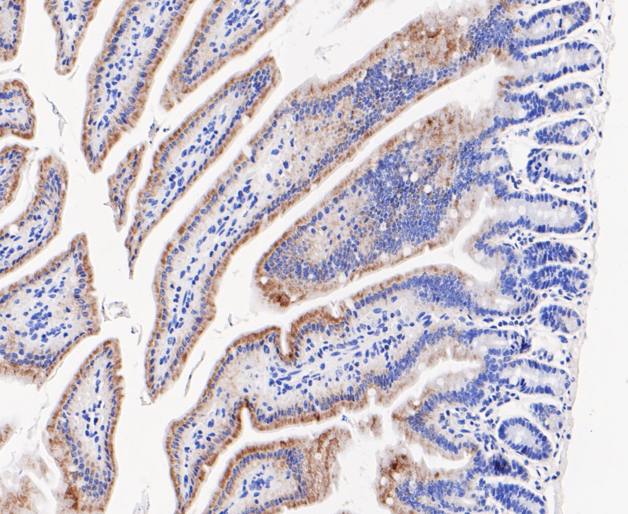
Fig5: Immunohistochemical analysis of paraffin-embedded mouse colon tissue using anti-ENTPD5 antibody. The section was pre-treated using heat mediated antigen retrieval with Tris-EDTA buffer (pH 8.0-8.4) for 20 minutes.The tissues were blocked in 5% BSA for 30 minutes at room temperature, washed with ddH2O and PBS, and then probed with the primary antibody (ET7109-86, 1/50) for 1 hour at room temperature. The detection was performed using an HRP conjugated compact polymer system. DAB was used as the chromogen. Tissues were counterstained with hematoxylin and mounted with DPX.Application
Fig5: Immunohistochemical analysis of paraffin-embedded mouse colon tissue using anti-ENTPD5 antibody. The section was pre-treated using heat mediated antigen retrieval with Tris-EDTA buffer (pH 8.0-8.4) for 20 minutes.The tissues were blocked in 5% BSA for 30 minutes at room temperature, washed with ddH2O and PBS, and then probed with the primary antibody (ET7109-86, 1/50) for 1 hour at room temperature. The detection was performed using an HRP conjugated compact polymer system. DAB was used as the chromogen. Tissues were counterstained with hematoxylin and mounted with DPX.Application
Fig5: Immunohistochemical analysis of paraffin-embedded mouse colon tissue using anti-ENTPD5 antibody. The section was pre-treated using heat mediated antigen retrieval with Tris-EDTA buffer (pH 8.0-8.4) for 20 minutes.The tissues were blocked in 5% BSA for 30 minutes at room temperature, washed with ddH2O and PBS, and then probed with the primary antibody (ET7109-86, 1/50) for 1 hour at room temperature. The detection was performed using an HRP conjugated compact polymer system. DAB was used as the chromogen. Tissues were counterstained with hematoxylin and mounted with DPX.Application
Fig5: Immunohistochemical analysis of paraffin-embedded mouse colon tissue using anti-ENTPD5 antibody. The section was pre-treated using heat mediated antigen retrieval with Tris-EDTA buffer (pH 8.0-8.4) for 20 minutes.The tissues were blocked in 5% BSA for 30 minutes at room temperature, washed with ddH2O and PBS, and then probed with the primary antibody (ET7109-86, 1/50) for 1 hour at room temperature. The detection was performed using an HRP conjugated compact polymer system. DAB was used as the chromogen. Tissues were counterstained with hematoxylin and mounted with DPX.Application
Fig5: Immunohistochemical analysis of paraffin-embedded mouse colon tissue using anti-ENTPD5 antibody. The section was pre-treated using heat mediated antigen retrieval with Tris-EDTA buffer (pH 8.0-8.4) for 20 minutes.The tissues were blocked in 5% BSA for 30 minutes at room temperature, washed with ddH2O and PBS, and then probed with the primary antibody (ET7109-86, 1/50) for 1 hour at room temperature. The detection was performed using an HRP conjugated compact polymer system. DAB was used as the chromogen. Tissues were counterstained with hematoxylin and mounted with DPX.| Product Name | ENTPD5 [JE48-94] |
|---|---|
| Antibody Type | Primary Antibodies |
| Antigen Alias | CD39 like 4 antibody AI196558 antibody AI987697 antibody CD39 antigen like 4 antibody CD39 antigen-like 4 antibody CD39 like 4 antibody CD39L4 antibody Ectonucleoside triphosphate diphosphohydrolase 5 antibody ENTP5_HUMAN antibody Entpd5 antibody ER UDPase antibody ER-UDPase antibody GDPase ENTPD5 antibody Guanosine-diphosphatase ENTPD5 antibody MGC163357 antibody MGC163359 antibody mNTPase antibody NTPDase 5 antibody Nucleoside diphosphatase antibody PCPH antibody Pcph proto oncogene protein antibody Proto oncogene CPH antibody UDPase ENTPD5 antibody Uridine-diphosphatase ENTPD5 antibody |
| Product description | Uridine diphosphatase (UDPase) that promotes protein N-glycosylation and ATP level regulation. UDP hydrolysis promotes protein N-glycosylation and folding in the endoplasmic reticulum, as well as elevated ATP consumption in the cytosol via an ATP hydrolysis cycle. Together with CMPK1 and AK1, constitutes an ATP hydrolysis cycle that converts ATP to AMP and results in a compensatory increase in aerobic glycolysis. The nucleotide hydrolyzing preference is GDP > IDP > UDP, but not any other nucleoside di-, mono- or triphosphates, nor thiamine pyrophosphate. Plays a key role in the AKT1-PTEN signaling pathway by promoting glycolysis in proliferating cells in response to phosphoinositide 3-kinase (PI3K) signaling. |
| Immunogen | Synthetic peptide within N-terminal human ENTPD5. |
| Clonality | Monoclonal |
|---|---|
| Isotype | IgG |
| Host Species | Recombinant rabbit |
| Tested Applications | WBIHCIP |
| WB:1:500-1:1,000: IP:1:50: IHC:1:50-1:200: |
|
| Species Reactivity | HumanMouseRat |
| Concentration | 1mg/ml |
| Alternative Names | CD39 like 4 antibody AI196558 antibody AI987697 antibody CD39 antigen like 4 antibody CD39 antigen-like 4 antibody CD39 like 4 antibody CD39L4 antibody Ectonucleoside triphosphate diphosphohydrolase 5 antibody ENTP5_HUMAN antibody Entpd5 antibody ER UDPase antibody ER-UDPase antibody GDPase ENTPD5 antibody Guanosine-diphosphatase ENTPD5 antibody MGC163357 antibody MGC163359 antibody mNTPase antibody NTPDase 5 antibody Nucleoside diphosphatase antibody PCPH antibody Pcph proto oncogene protein antibody Proto oncogene CPH antibody UDPase ENTPD5 antibody Uridine-diphosphatase ENTPD5 antibody |
|---|---|
| Molecular Weight(MW) | 47 kDa |
| Cellular Localization | Endoplasmic reticulum. Secreted. |
Application
Fig5: Immunohistochemical analysis of paraffin-embedded mouse colon tissue using anti-ENTPD5 antibody. The section was pre-treated using heat mediated antigen retrieval with Tris-EDTA buffer (pH 8.0-8.4) for 20 minutes.The tissues were blocked in 5% BSA for 30 minutes at room temperature, washed with ddH2O and PBS, and then probed with the primary antibody (ET7109-86, 1/50) for 1 hour at room temperature. The detection was performed using an HRP conjugated compact polymer system. DAB was used as the chromogen. Tissues were counterstained with hematoxylin and mounted with DPX.
Application
Fig5: Immunohistochemical analysis of paraffin-embedded mouse colon tissue using anti-ENTPD5 antibody. The section was pre-treated using heat mediated antigen retrieval with Tris-EDTA buffer (pH 8.0-8.4) for 20 minutes.The tissues were blocked in 5% BSA for 30 minutes at room temperature, washed with ddH2O and PBS, and then probed with the primary antibody (ET7109-86, 1/50) for 1 hour at room temperature. The detection was performed using an HRP conjugated compact polymer system. DAB was used as the chromogen. Tissues were counterstained with hematoxylin and mounted with DPX.
Application
Fig5: Immunohistochemical analysis of paraffin-embedded mouse colon tissue using anti-ENTPD5 antibody. The section was pre-treated using heat mediated antigen retrieval with Tris-EDTA buffer (pH 8.0-8.4) for 20 minutes.The tissues were blocked in 5% BSA for 30 minutes at room temperature, washed with ddH2O and PBS, and then probed with the primary antibody (ET7109-86, 1/50) for 1 hour at room temperature. The detection was performed using an HRP conjugated compact polymer system. DAB was used as the chromogen. Tissues were counterstained with hematoxylin and mounted with DPX.
Application
Fig5: Immunohistochemical analysis of paraffin-embedded mouse colon tissue using anti-ENTPD5 antibody. The section was pre-treated using heat mediated antigen retrieval with Tris-EDTA buffer (pH 8.0-8.4) for 20 minutes.The tissues were blocked in 5% BSA for 30 minutes at room temperature, washed with ddH2O and PBS, and then probed with the primary antibody (ET7109-86, 1/50) for 1 hour at room temperature. The detection was performed using an HRP conjugated compact polymer system. DAB was used as the chromogen. Tissues were counterstained with hematoxylin and mounted with DPX.
Application
Fig5: Immunohistochemical analysis of paraffin-embedded mouse colon tissue using anti-ENTPD5 antibody. The section was pre-treated using heat mediated antigen retrieval with Tris-EDTA buffer (pH 8.0-8.4) for 20 minutes.The tissues were blocked in 5% BSA for 30 minutes at room temperature, washed with ddH2O and PBS, and then probed with the primary antibody (ET7109-86, 1/50) for 1 hour at room temperature. The detection was performed using an HRP conjugated compact polymer system. DAB was used as the chromogen. Tissues were counterstained with hematoxylin and mounted with DPX.| Positive Control | Human liver tissue, rat testis tissue, human liver cancer tissue, human kidney tissue, mouse colon tissue. |
|---|---|
| Application Notes | WB:1:500-1:1,000: IP:1:50: IHC:1:50-1:200: |
| Form | Liquid |
|---|---|
| Storage Instructions | Store at +4℃ after thawing. Aliquot store at -20℃. Avoid repeated freeze / thaw cycles. |
| Storage Buffer | 1*TBS (pH7.4), 1%BSA, 50%Glycerol. Preservative: 0.05% Sodium Azide. |


