Application
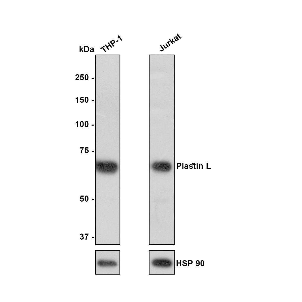
Fig1: Western blot analysis of Plastin L on different lysates with Rabbit anti-Plastin L antibody at 1/500 dilution. Lane 1: THP-1 (Human acute monoblastic leukemia cell) cell lysate, Lane 2: Jurkat (Human T-lymphoblastic cells) cell lysate.Application
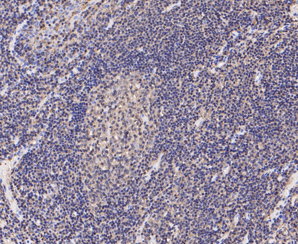
Fig2: Immunohistochemical analysis of paraffin-embedded human tonsil tissue using anti-Plastin L antibody. The section was pre-treated using heat mediated antigen retrieval with Tris-EDTA buffer (pH 8.0-8.4) for 20 minutes.The tissues were blocked in 5% BSA for 30 minutes at room temperature, washed with ddH2O and PBS, and then probed with the primary antibody (1/200) for 30 minutes at room temperature. The detection was performed using an HRP conjugated compact polymer system. DAB was used as the chromogen. Tissues were counterstained with hematoxylin and mounted with DPX.Application
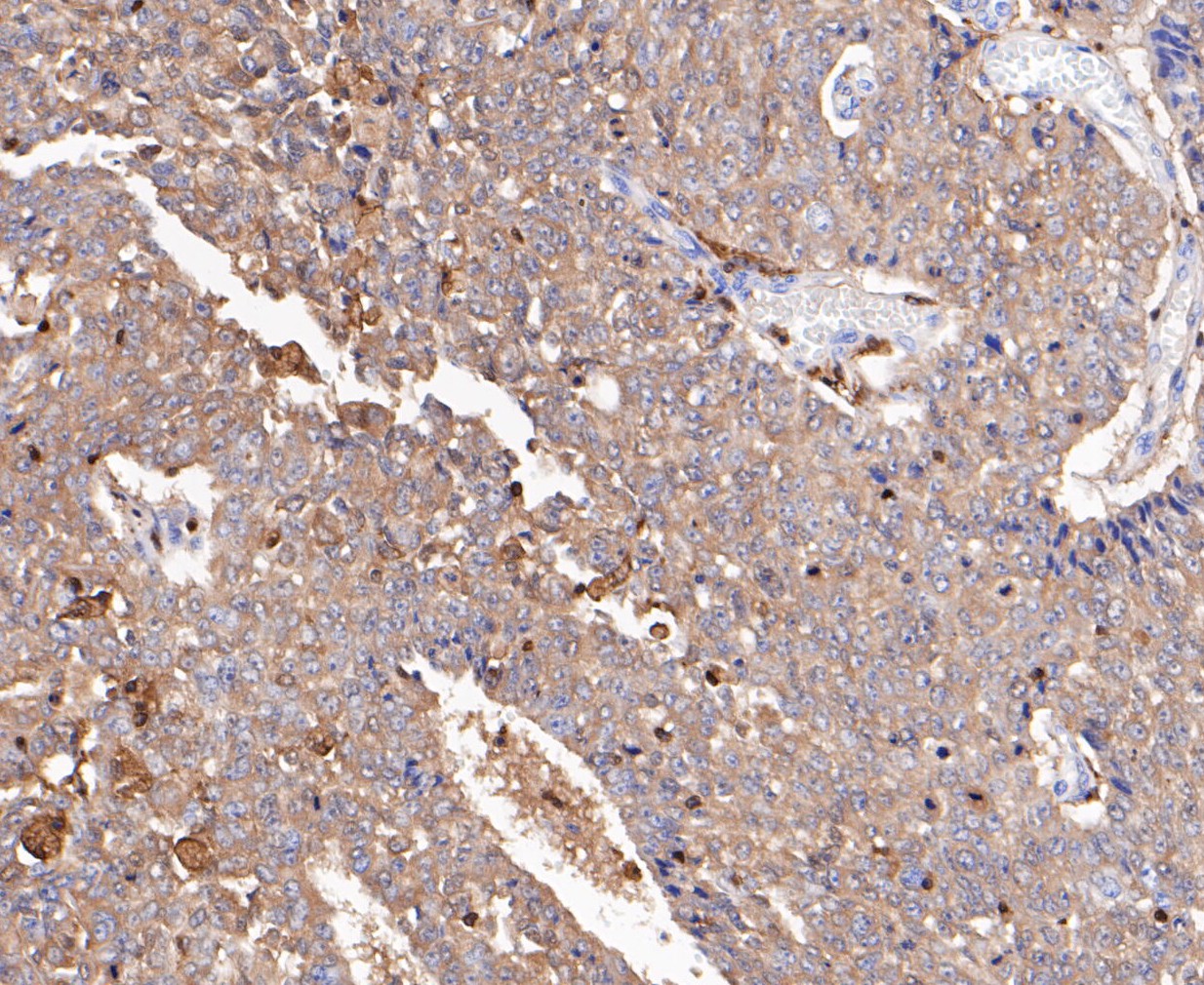
Fig3: Immunohistochemical analysis of paraffin-embedded human lung cancer tissue using anti-Plastin L antibody. The section was pre-treated using heat mediated antigen retrieval with Tris-EDTA buffer (pH 8.0-8.4) for 20 minutes.The tissues were blocked in 5% BSA for 30 minutes at room temperature, washed with ddH2O and PBS, and then probed with the primary antibody (1/200) for 30 minutes at room temperature. The detection was performed using an HRP conjugated compact polymer system. DAB was used as the chromogen. Tissues were counterstained with hematoxylin and mounted with DPX.Application
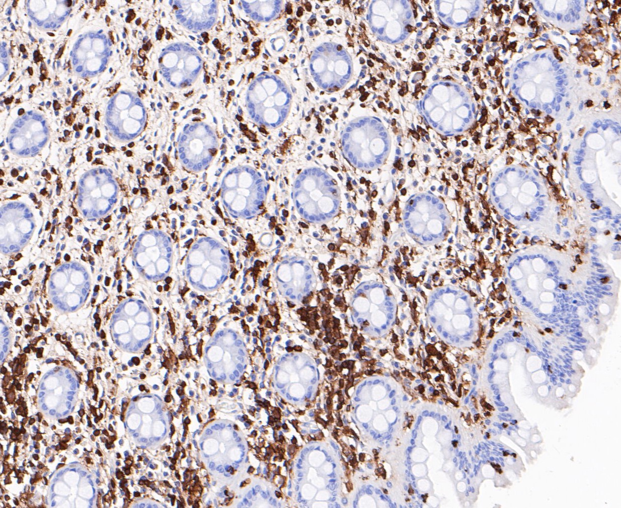
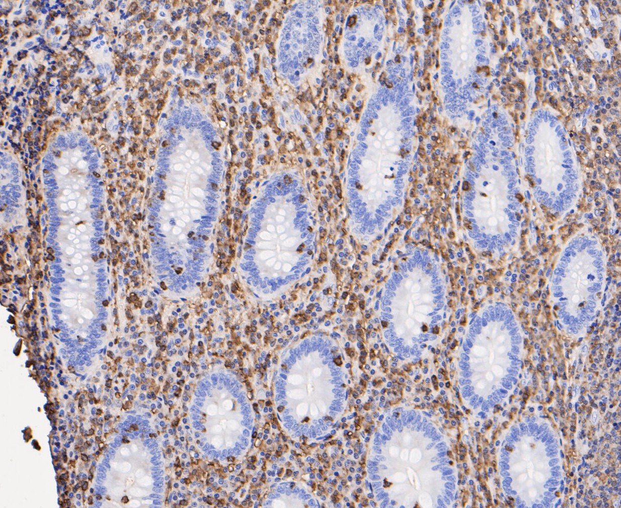
Fig4: Immunohistochemical analysis of paraffin-embedded human colon tissue using anti-Plastin L antibody. The section was pre-treated using heat mediated antigen retrieval with Tris-EDTA buffer (pH 8.0-8.4) for 20 minutes.The tissues were blocked in 5% BSA for 30 minutes at room temperature, washed with ddH2O and PBS, and then probed with the primary antibody (1/200) for 30 minutes at room temperature. The detection was performed using an HRP conjugated compact polymer system. DAB was used as the chromogen. Tissues were counterstained with hematoxylin and mounted with DPX.Application
Fig5: Immunohistochemical analysis of paraffin-embedded human appendix tissue using anti-Plastin L antibody. The section was pre-treated using heat mediated antigen retrieval with Tris-EDTA buffer (pH 8.0-8.4) for 20 minutes.The tissues were blocked in 5% BSA for 30 minutes at room temperature, washed with ddH2O and PBS, and then probed with the primary antibody (1/200) for 30 minutes at room temperature. The detection was performed using an HRP conjugated compact polymer system. DAB was used as the chromogen. Tissues were counterstained with hematoxylin and mounted with DPX.Application
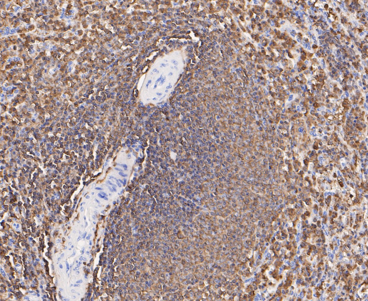
Fig6: Immunohistochemical analysis of paraffin-embedded human spleen tissue using anti-Plastin L antibody. The section was pre-treated using heat mediated antigen retrieval with Tris-EDTA buffer (pH 8.0-8.4) for 20 minutes.The tissues were blocked in 5% BSA for 30 minutes at room temperature, washed with ddH2O and PBS, and then probed with the primary antibody (1/50) for 30 minutes at room temperature. The detection was performed using an HRP conjugated compact polymer system. DAB was used as the chromogen. Tissues were counterstained with hematoxylin and mounted with DPX.Application
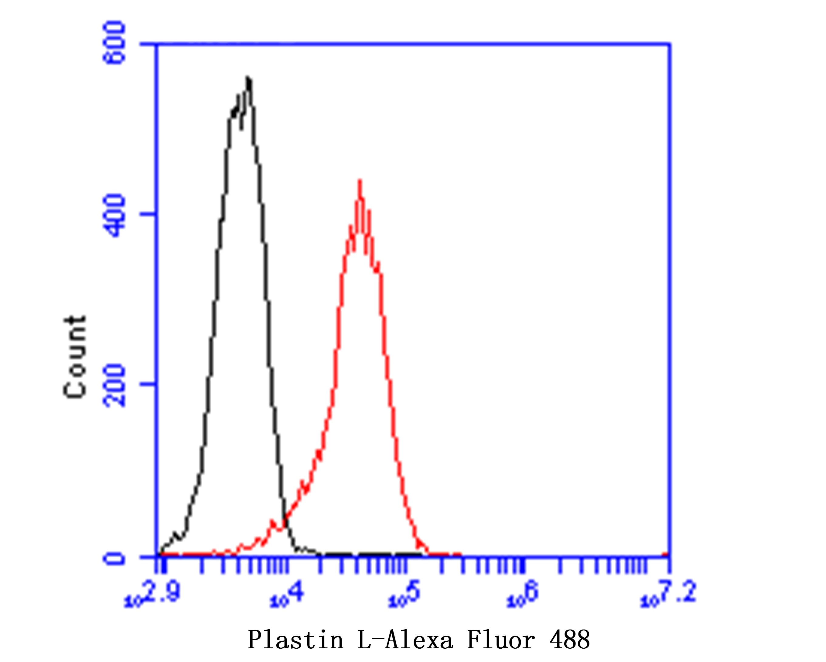
Fig7: Flow cytometric analysis of Plastin L was done on THP-1 cells. The cells were fixed, permeabilized and stained with Plastin L antibody at 1/100 dilution (red) compared with an unlabelled control (cells without incubation with primary antibody; black). After incubation of the primary antibody on room temperature for an hour, the cells was stained with a Alexa Fluor™ 488-conjugated goat anti-rabbit IgG Secondary antibody at 1/500 dilution for 30 minutes.| Product Name | Plastin L Recombinant Rabbit Monoclonal Antibody |
|---|---|
| Antibody Type | Primary Antibodies |
| Product description | Plastins are a family of actin-binding proteins that are conserved throughout eukaryote evolution and expressed in most tissues of higher eukaryotes. In humans, two ubiquitous plastin isoforms (L and T) have been identified. Plastin 1 (otherwise known as Fimbrin) is a third distinct plastin isoform which is specifically expressed at high levels in the small intestine. The L isoform is expressed only in hemopoietic cell lineages, while the T isoform has been found in all other normal cells of solid tissues that have replicative potential (fibroblasts, endothelial cells, epithelial cells, melanocytes, etc.). However, L-plastin has been found in many types of malignant human cells of non-hemopoietic origin suggesting that its expression is induced accompanying tumorigenesis in solid tissues. |
| Immunogen | Synthetic peptide within C terminal human Plastin L. |
| Clonality | Monoclonal |
|---|---|
| Isotype | IgG |
| Host Species | Recombinant rabbit |
| Tested Applications | FCIHCWB |
| WB:1:500-1:2,000: IHC:1:50-1:200: FC:1:50-1:100: |
|
| Species Reactivity | Human |
| Concentration | 1mg/ml |
| Purification | Protein A |
| Alternative Names | CP64 antibody L plastin antibody L-plastin antibody Larval cuticle protein 1 antibody LC64P antibody LCP-1 antibody LCP1 antibody LPL antibody Lplastin antibody Lymphocyte cytosolic protein 1 antibody Plastin 2 antibody Plastin-2 antibody PLS2 antibody PLSL_HUMAN antibody |
|---|---|
| Molecular Weight(MW) | 70 kDa |
| Cellular Localization | Plasma membrane. Cytoskeleton. |
| SwissProt ID | P13796 |
|---|
Application
Fig1: Western blot analysis of Plastin L on different lysates with Rabbit anti-Plastin L antibody at 1/500 dilution. Lane 1: THP-1 (Human acute monoblastic leukemia cell) cell lysate, Lane 2: Jurkat (Human T-lymphoblastic cells) cell lysate.
Application
Fig2: Immunohistochemical analysis of paraffin-embedded human tonsil tissue using anti-Plastin L antibody. The section was pre-treated using heat mediated antigen retrieval with Tris-EDTA buffer (pH 8.0-8.4) for 20 minutes.The tissues were blocked in 5% BSA for 30 minutes at room temperature, washed with ddH2O and PBS, and then probed with the primary antibody (1/200) for 30 minutes at room temperature. The detection was performed using an HRP conjugated compact polymer system. DAB was used as the chromogen. Tissues were counterstained with hematoxylin and mounted with DPX.
Application
Fig3: Immunohistochemical analysis of paraffin-embedded human lung cancer tissue using anti-Plastin L antibody. The section was pre-treated using heat mediated antigen retrieval with Tris-EDTA buffer (pH 8.0-8.4) for 20 minutes.The tissues were blocked in 5% BSA for 30 minutes at room temperature, washed with ddH2O and PBS, and then probed with the primary antibody (1/200) for 30 minutes at room temperature. The detection was performed using an HRP conjugated compact polymer system. DAB was used as the chromogen. Tissues were counterstained with hematoxylin and mounted with DPX.
Application
Fig4: Immunohistochemical analysis of paraffin-embedded human colon tissue using anti-Plastin L antibody. The section was pre-treated using heat mediated antigen retrieval with Tris-EDTA buffer (pH 8.0-8.4) for 20 minutes.The tissues were blocked in 5% BSA for 30 minutes at room temperature, washed with ddH2O and PBS, and then probed with the primary antibody (1/200) for 30 minutes at room temperature. The detection was performed using an HRP conjugated compact polymer system. DAB was used as the chromogen. Tissues were counterstained with hematoxylin and mounted with DPX.
Application
Fig5: Immunohistochemical analysis of paraffin-embedded human appendix tissue using anti-Plastin L antibody. The section was pre-treated using heat mediated antigen retrieval with Tris-EDTA buffer (pH 8.0-8.4) for 20 minutes.The tissues were blocked in 5% BSA for 30 minutes at room temperature, washed with ddH2O and PBS, and then probed with the primary antibody (1/200) for 30 minutes at room temperature. The detection was performed using an HRP conjugated compact polymer system. DAB was used as the chromogen. Tissues were counterstained with hematoxylin and mounted with DPX.
Application
Fig6: Immunohistochemical analysis of paraffin-embedded human spleen tissue using anti-Plastin L antibody. The section was pre-treated using heat mediated antigen retrieval with Tris-EDTA buffer (pH 8.0-8.4) for 20 minutes.The tissues were blocked in 5% BSA for 30 minutes at room temperature, washed with ddH2O and PBS, and then probed with the primary antibody (1/50) for 30 minutes at room temperature. The detection was performed using an HRP conjugated compact polymer system. DAB was used as the chromogen. Tissues were counterstained with hematoxylin and mounted with DPX.
Application
Fig7: Flow cytometric analysis of Plastin L was done on THP-1 cells. The cells were fixed, permeabilized and stained with Plastin L antibody at 1/100 dilution (red) compared with an unlabelled control (cells without incubation with primary antibody; black). After incubation of the primary antibody on room temperature for an hour, the cells was stained with a Alexa Fluor™ 488-conjugated goat anti-rabbit IgG Secondary antibody at 1/500 dilution for 30 minutes.| Positive Control | Human tonsil tissue, human lung cancer tissue, human colon tissue, human appendix tissue, human spleen tissue. |
|---|---|
| Application Notes | WB:1:500-1:2,000: IHC:1:50-1:200: FC:1:50-1:100: |
| Form | Liquid |
|---|---|
| Storage Instructions | Store at +4℃ after thawing. Aliquot store at -20℃. Avoid repeated freeze / thaw cycles. |
| Storage Buffer | 1*TBS (pH7.4), 1%BSA, 50%Glycerol. Preservative: 0.05% Sodium Azide. |


