Application
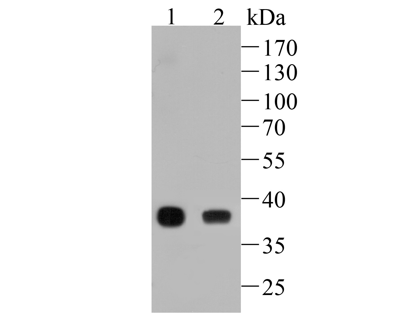
Fig1: Western blot analysis of FBP1 on different lysates. Proteins were transferred to a PVDF membrane and blocked with 5% BSA in PBS for 1 hour at room temperature. The primary antibody (ET7110-07, 1/500) was used in 5% BSA at room temperature for 2 hours. Goat Anti-Rabbit IgG - HRP Secondary Antibody (HA1001) at 1:5,000 dilution was used for 1 hour at room temperature. Positive control: Lane 1: Bouse liver tissue lysate Lane 2: Rat liver tissue lysateApplication
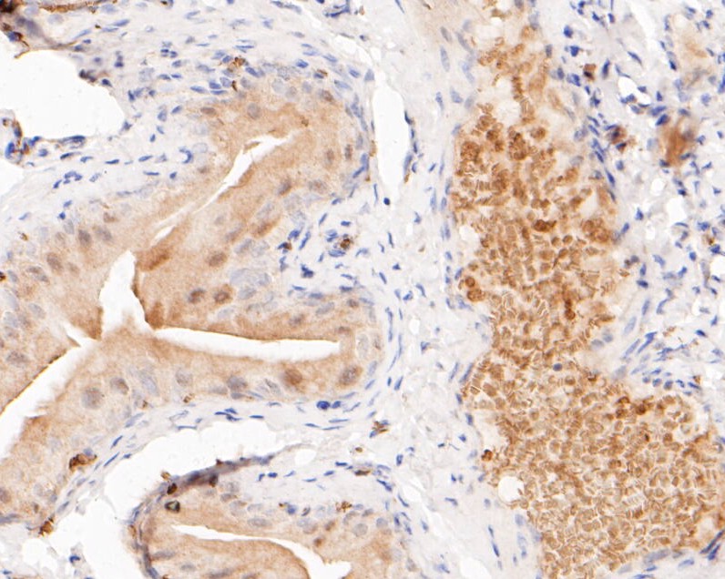
Fig2: Immunohistochemical analysis of paraffin-embedded rat bladder tissue using anti-FBP1 antibody. The section was pre-treated using heat mediated antigen retrieval with sodium citrate buffer (pH 6.0) for 20 minutes. The tissues were blocked in 5% BSA for 30 minutes at room temperature, washed with ddH2O and PBS, and then probed with the primary antibody (ET7110-07, 1/200) for 30 minutes at room temperature. The detection was performed using an HRP conjugated compact polymer system. DAB was used as the chromogen. Tissues were counterstained with hematoxylin and mounted with DPX.Application
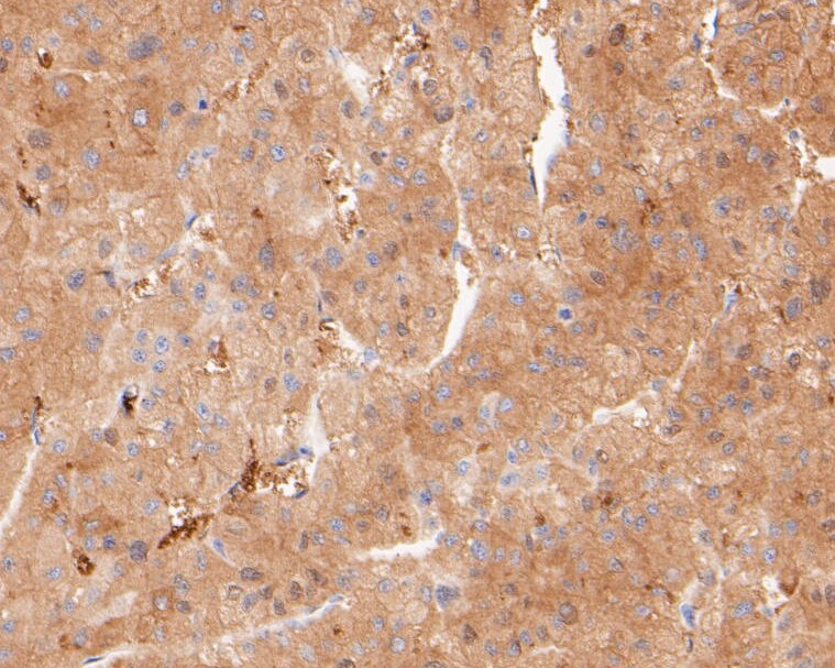
Fig3: Immunohistochemical analysis of paraffin-embedded human liver tissue using anti-FBP1 antibody. The section was pre-treated using heat mediated antigen retrieval with sodium citrate buffer (pH 6.0) for 20 minutes. The tissues were blocked in 5% BSA for 30 minutes at room temperature, washed with ddH2O and PBS, and then probed with the primary antibody (ET7110-07, 1/200) for 30 minutes at room temperature. The detection was performed using an HRP conjugated compact polymer system. DAB was used as the chromogen. Tissues were counterstained with hematoxylin and mounted with DPX.Application
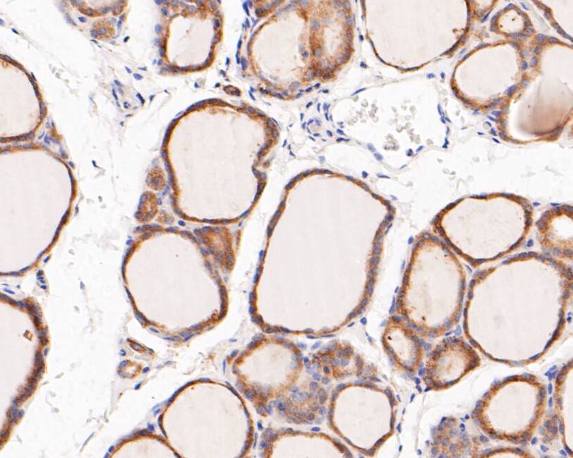
Fig4: Immunohistochemical analysis of paraffin-embedded human thyroid carcinoma tissue using anti-FBP1 antibody. The section was pre-treated using heat mediated antigen retrieval with sodium citrate buffer (pH 6.0) for 20 minutes. The tissues were blocked in 5% BSA for 30 minutes at room temperature, washed with ddH2O and PBS, and then probed with the primary antibody (ET7110-07, 1/200) for 30 minutes at room temperature. The detection was performed using an HRP conjugated compact polymer system. DAB was used as the chromogen. Tissues were counterstained with hematoxylin and mounted with DPX.Application
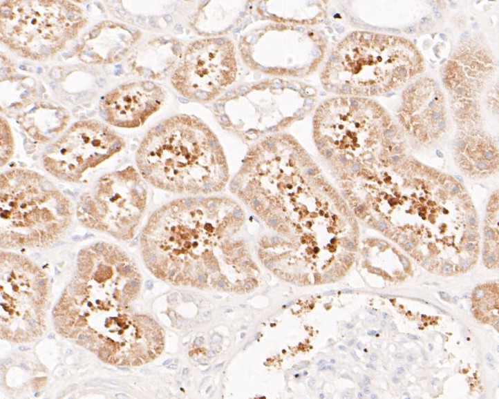
Fig5: Immunohistochemical analysis of paraffin-embedded human kidney tissue using anti-FBP1 antibody. The section was pre-treated using heat mediated antigen retrieval with sodium citrate buffer (pH 6.0) for 20 minutes. The tissues were blocked in 5% BSA for 30 minutes at room temperature, washed with ddH2O and PBS, and then probed with the primary antibody (ET7110-07, 1/200) for 30 minutes at room temperature. The detection was performed using an HRP conjugated compact polymer system. DAB was used as the chromogen. Tissues were counterstained with hematoxylin and mounted with DPX.Application
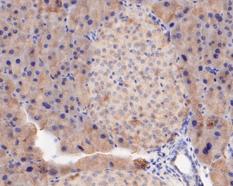
Fig6: Immunohistochemical analysis of paraffin-embedded mouse pancreas tissue using anti-FBP1 antibody. The section was pre-treated using heat mediated antigen retrieval with sodium citrate buffer (pH 6.0) for 20 minutes. The tissues were blocked in 5% BSA for 30 minutes at room temperature, washed with ddH2O and PBS, and then probed with the primary antibody (ET7110-07, 1/200) for 30 minutes at room temperature. The detection was performed using an HRP conjugated compact polymer system. DAB was used as the chromogen. Tissues were counterstained with hematoxylin and mounted with DPX.| Product Name | FBP1 [JE50-76] |
|---|---|
| Antibody Type | Primary Antibodies |
| Antigen Alias | 6-bisphosphatase 1 antibody 6-bisphosphate 1-phosphohydrolase 1 antibody D fructose 1 6 bisphosphate 1 phosphohydrolase 1 antibody D-fructose-1 antibody EC 3.1.3.11 antibody F16P1_HUMAN antibody FBP antibody FBP 1 antibody FBP1 antibody FBPase 1 antibody Fructose 1 6 bisphosphatase 1 antibody Fructose bisphosphatase 1 antibody Fructose-1 antibody Growth inhibiting protein 17 antibody Liver fructose bisphosphatase antibody |
| Product description | Fructose-1,6-bisphosphatase 1 is a protein that in humans is encoded by the FBP1 gene. Fructose-1,6-bisphosphatase 1, catalyzes the hydrolysis of fructose 1,6-bisphosphate to fructose 6-phosphate in the presence of divalent cations, acting as a rate-limiting enzyme in gluconeogenesis. Plays a role in regulating glucose sensing and insulin secretion of pancreatic beta-cells. Appears to modulate glycerol gluconeogenesis in liver. Important regulator of appetite and adiposity; increased expression of the protein in liver after nutrient excess increases circulating satiety hormones and reduces appetite-stimulating neuropeptides and thus seems to provide a feedback mechanism to limit weight gain. Fructose-1,6-diphosphatase deficiency is associated with hypoglycemia and metabolic acidosis. |
| Immunogen | Synthetic peptide within N-terminal human FBP1 |
| Clonality | Polyclonal |
|---|---|
| Isotype | IgG |
| Host Species | Rabbit |
| Tested Applications | WBIHC-P |
| WB:1:500-1:2,000 IHC-P:1:50-1:200 |
|
| Species Reactivity | HumanMouseRat |
| Concentration | 1mg/ml |
| Alternative Names | 6-bisphosphatase 1 antibody
6-bisphosphate 1-phosphohydrolase 1 antibody
D fructose 1 6 bisphosphate 1 phosphohydrolase 1 antibody D-fructose-1 antibody EC 3.1.3.11 antibody F16P1_HUMAN antibody FBP antibody FBP 1 antibody FBP1 antibody FBPase 1 antibody Fructose 1 6 bisphosphatase 1 antibody Fructose bisphosphatase 1 antibody Fructose-1 antibody Growth inhibiting protein 17 antibody Liver fructose bisphosphatase antibody |
|---|---|
| Molecular Weight(MW) | 37 kDa |
| Cellular Localization | Cytosol, extracellular exosome, nucleus, cytoplasm. |
Application
Fig1: Western blot analysis of FBP1 on different lysates. Proteins were transferred to a PVDF membrane and blocked with 5% BSA in PBS for 1 hour at room temperature. The primary antibody (ET7110-07, 1/500) was used in 5% BSA at room temperature for 2 hours. Goat Anti-Rabbit IgG - HRP Secondary Antibody (HA1001) at 1:5,000 dilution was used for 1 hour at room temperature. Positive control: Lane 1: Bouse liver tissue lysate Lane 2: Rat liver tissue lysate
Application
Fig2: Immunohistochemical analysis of paraffin-embedded rat bladder tissue using anti-FBP1 antibody. The section was pre-treated using heat mediated antigen retrieval with sodium citrate buffer (pH 6.0) for 20 minutes. The tissues were blocked in 5% BSA for 30 minutes at room temperature, washed with ddH2O and PBS, and then probed with the primary antibody (ET7110-07, 1/200) for 30 minutes at room temperature. The detection was performed using an HRP conjugated compact polymer system. DAB was used as the chromogen. Tissues were counterstained with hematoxylin and mounted with DPX.
Application
Fig3: Immunohistochemical analysis of paraffin-embedded human liver tissue using anti-FBP1 antibody. The section was pre-treated using heat mediated antigen retrieval with sodium citrate buffer (pH 6.0) for 20 minutes. The tissues were blocked in 5% BSA for 30 minutes at room temperature, washed with ddH2O and PBS, and then probed with the primary antibody (ET7110-07, 1/200) for 30 minutes at room temperature. The detection was performed using an HRP conjugated compact polymer system. DAB was used as the chromogen. Tissues were counterstained with hematoxylin and mounted with DPX.
Application
Fig4: Immunohistochemical analysis of paraffin-embedded human thyroid carcinoma tissue using anti-FBP1 antibody. The section was pre-treated using heat mediated antigen retrieval with sodium citrate buffer (pH 6.0) for 20 minutes. The tissues were blocked in 5% BSA for 30 minutes at room temperature, washed with ddH2O and PBS, and then probed with the primary antibody (ET7110-07, 1/200) for 30 minutes at room temperature. The detection was performed using an HRP conjugated compact polymer system. DAB was used as the chromogen. Tissues were counterstained with hematoxylin and mounted with DPX.
Application
Fig5: Immunohistochemical analysis of paraffin-embedded human kidney tissue using anti-FBP1 antibody. The section was pre-treated using heat mediated antigen retrieval with sodium citrate buffer (pH 6.0) for 20 minutes. The tissues were blocked in 5% BSA for 30 minutes at room temperature, washed with ddH2O and PBS, and then probed with the primary antibody (ET7110-07, 1/200) for 30 minutes at room temperature. The detection was performed using an HRP conjugated compact polymer system. DAB was used as the chromogen. Tissues were counterstained with hematoxylin and mounted with DPX.
Application
Fig6: Immunohistochemical analysis of paraffin-embedded mouse pancreas tissue using anti-FBP1 antibody. The section was pre-treated using heat mediated antigen retrieval with sodium citrate buffer (pH 6.0) for 20 minutes. The tissues were blocked in 5% BSA for 30 minutes at room temperature, washed with ddH2O and PBS, and then probed with the primary antibody (ET7110-07, 1/200) for 30 minutes at room temperature. The detection was performed using an HRP conjugated compact polymer system. DAB was used as the chromogen. Tissues were counterstained with hematoxylin and mounted with DPX.| Positive Control | Mouse liver tissue, rat liver tissue, rat bladder tissue, human liver tissue, human thyroid carcinoma tissue, human kidney tissue, mouse pancreas tissue. |
|---|---|
| Application Notes | WB:1:500-1:2,000 IHC-P:1:50-1:200 |
| Form | Liquid |
|---|---|
| Storage Instructions | Store at +4℃ after thawing. Aliquot store at -20℃. Avoid repeated freeze / thaw cycles. |
| Storage Buffer | 1*TBS (pH7.4), 1%BSA, 50%Glycerol. Preservative: 0.05% Sodium Azide. |


