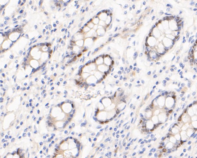
Application
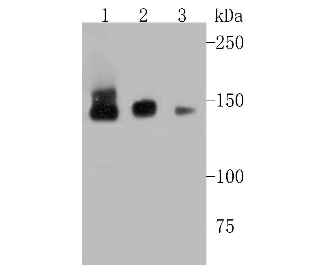
Fig1: Western blot analysis of Timeless on different lysates. Proteins were transferred to a PVDF membrane and blocked with 5% BSA in PBS for 1 hour at room temperature. The primary antibody (ET7110-23, 1/500) was used in 5% BSA at room temperature for 2 hours. Goat Anti-Rabbit IgG - HRP Secondary Antibody (HA1001) at 1:5,000 dilution was used for 1 hour at room temperature. Positive control: Lane 1: Hela cell lysate Lane 2: 293T cell lysate Lane 3: Daudi cell lysateApplication
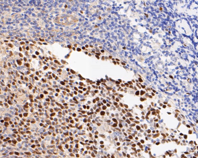
Fig2: Immunohistochemical analysis of paraffin-embedded human tonsil tissue using anti-Timeless antibody. The section was pre-treated using heat mediated antigen retrieval with sodium citrate buffer (pH 6.0) for 20 minutes. The tissues were blocked in 5% BSA for 30 minutes at room temperature, washed with ddH2O and PBS, and then probed with the primary antibody (ET7110-23, 1/50) for 30 minutes at room temperature. The detection was performed using an HRP conjugated compact polymer system. DAB was used as the chromogen. Tissues were counterstained with hematoxylin and mounted with DPX.Application
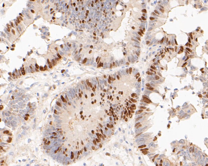
Fig3: Immunohistochemical analysis of paraffin-embedded human colon carcinoma tissue using anti-Timeless antibody. The section was pre-treated using heat mediated antigen retrieval with sodium citrate buffer (pH 6.0) for 20 minutes. The tissues were blocked in 5% BSA for 30 minutes at room temperature, washed with ddH2O and PBS, and then probed with the primary antibody (ET7110-23, 1/50) for 30 minutes at room temperature. The detection was performed using an HRP conjugated compact polymer system. DAB was used as the chromogen. Tissues were counterstained with hematoxylin and mounted with DPX.Application
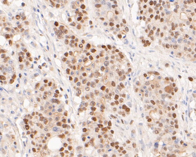
Fig4: Immunohistochemical analysis of paraffin-embedded human gastric carcinoma tissue using anti-Timeless antibody. The section was pre-treated using heat mediated antigen retrieval with sodium citrate buffer (pH 6.0) for 20 minutes. The tissues were blocked in 5% BSA for 30 minutes at room temperature, washed with ddH2O and PBS, and then probed with the primary antibody (ET7110-23, 1/50) for 30 minutes at room temperature. The detection was performed using an HRP conjugated compact polymer system. DAB was used as the chromogen. Tissues were counterstained with hematoxylin and mounted with DPX.Application
Fig5: Immunohistochemical analysis of paraffin-embedded human small intestine tissue using anti-Timeless antibody. The section was pre-treated using heat mediated antigen retrieval with sodium citrate buffer (pH 6.0) for 20 minutes. The tissues were blocked in 5% BSA for 30 minutes at room temperature, washed with ddH2O and PBS, and then probed with the primary antibody (ET7110-23, 1/50) for 30 minutes at room temperature. The detection was performed using an HRP conjugated compact polymer system. DAB was used as the chromogen. Tissues were counterstained with hematoxylin and mounted with DPX.| Product Name | Timeless [JE51-39] |
|---|---|
| Antibody Type | Primary Antibodies |
| Antigen Alias | FLJ12640 antibody FLJ20714 antibody hTIM antibody Protein timeless homolog antibody TIM antibody TIM_HUMAN antibody TIM1 antibody Timeless antibody timeless circadian clock 1 antibody timeless circadian clock antibody timeless homolog antibody TIMELESS1 antibody Tof1 homolog antibody |
| Product description | Plays an important role in the control of DNA replication, maintenance of replication fork stability, maintenance of genome stability throughout normal DNA replication, DNA repair and in the regulation of the circadian clock. Required to stabilize replication forks during DNA replication by forming a complex with TIPIN: this complex regulates DNA replication processes under both normal and stress conditions, stabilizes replication forks and influences both CHEK1 phosphorylation and the intra-S phase checkpoint in response to genotoxic stress. TIMELESS promotes TIPIN nuclear localization. Involved in cell survival after DNA damage or replication stress by promoting DNA repair. In response to double-strand breaks (DSBs), accumulates at DNA damage sites and promotes homologous recombination repair via its interaction with PARP1. May be specifically required for the ATR-CHEK1 pathway in the replication checkpoint induced by hydroxyurea or ultraviolet light. Involved in the determination of period length and in the DNA damage-dependent phase advancing of the circadian clock. Negatively regulates CLOCK|NPAS2-ARTNL/BMAL1|ARTNL2/BMAL2-induced transactivation of PER1 possibly via translocation of PER1 into the nucleus. May also play an important role in epithelial cell morphogenesis and formation of branching tubules (By similarity). |
| Immunogen | Synthetic peptide within C-terminal human Timeless |
| Clonality | Monoclonal |
|---|---|
| Isotype | IgG |
| Host Species | Recombinant rabbit |
| Tested Applications | WBIHC-P |
| WB:1:500-1:2,000 IHC-P:1:50-1:200 |
|
| Species Reactivity | Human |
| Concentration | 1mg/mL |
| Alternative Names | FLJ12640 antibody
FLJ20714 antibody
hTIM antibody Protein timeless homolog antibody TIM antibody TIM_HUMAN antibody TIM1 antibody Timeless antibody timeless circadian clock 1 antibody timeless circadian clock antibody timeless homolog antibody TIMELESS1 antibody Tof1 homolog antibody |
|---|---|
| Molecular Weight(MW) | 139 kDa |
| Cellular Localization | Nucleus, chromosome. |
| SwissProt ID | Q9UNS1 |
|---|
Application
Fig1: Western blot analysis of Timeless on different lysates. Proteins were transferred to a PVDF membrane and blocked with 5% BSA in PBS for 1 hour at room temperature. The primary antibody (ET7110-23, 1/500) was used in 5% BSA at room temperature for 2 hours. Goat Anti-Rabbit IgG - HRP Secondary Antibody (HA1001) at 1:5,000 dilution was used for 1 hour at room temperature. Positive control: Lane 1: Hela cell lysate Lane 2: 293T cell lysate Lane 3: Daudi cell lysate
Application
Fig2: Immunohistochemical analysis of paraffin-embedded human tonsil tissue using anti-Timeless antibody. The section was pre-treated using heat mediated antigen retrieval with sodium citrate buffer (pH 6.0) for 20 minutes. The tissues were blocked in 5% BSA for 30 minutes at room temperature, washed with ddH2O and PBS, and then probed with the primary antibody (ET7110-23, 1/50) for 30 minutes at room temperature. The detection was performed using an HRP conjugated compact polymer system. DAB was used as the chromogen. Tissues were counterstained with hematoxylin and mounted with DPX.
Application
Fig3: Immunohistochemical analysis of paraffin-embedded human colon carcinoma tissue using anti-Timeless antibody. The section was pre-treated using heat mediated antigen retrieval with sodium citrate buffer (pH 6.0) for 20 minutes. The tissues were blocked in 5% BSA for 30 minutes at room temperature, washed with ddH2O and PBS, and then probed with the primary antibody (ET7110-23, 1/50) for 30 minutes at room temperature. The detection was performed using an HRP conjugated compact polymer system. DAB was used as the chromogen. Tissues were counterstained with hematoxylin and mounted with DPX.
Application
Fig4: Immunohistochemical analysis of paraffin-embedded human gastric carcinoma tissue using anti-Timeless antibody. The section was pre-treated using heat mediated antigen retrieval with sodium citrate buffer (pH 6.0) for 20 minutes. The tissues were blocked in 5% BSA for 30 minutes at room temperature, washed with ddH2O and PBS, and then probed with the primary antibody (ET7110-23, 1/50) for 30 minutes at room temperature. The detection was performed using an HRP conjugated compact polymer system. DAB was used as the chromogen. Tissues were counterstained with hematoxylin and mounted with DPX.
Application
Fig5: Immunohistochemical analysis of paraffin-embedded human small intestine tissue using anti-Timeless antibody. The section was pre-treated using heat mediated antigen retrieval with sodium citrate buffer (pH 6.0) for 20 minutes. The tissues were blocked in 5% BSA for 30 minutes at room temperature, washed with ddH2O and PBS, and then probed with the primary antibody (ET7110-23, 1/50) for 30 minutes at room temperature. The detection was performed using an HRP conjugated compact polymer system. DAB was used as the chromogen. Tissues were counterstained with hematoxylin and mounted with DPX.| Positive Control | Hela cell lysates, 293T cell lysates, Daudi cell lysates, human tonsil tissue, human colon carcinoma tissue, human gastric carcinoma tissue, human small intestine tissue, human pancreas tissue. |
|---|---|
| Application Notes | WB:1:500-1:2,000 IHC-P:1:50-1:200 |
| Form | Liquid |
|---|---|
| Storage Instructions | Store at +4℃ after thawing. Aliquot store at -20℃. Avoid repeated freeze / thaw cycles. |
| Storage Buffer | 1*TBS (pH7.4), 1%BSA, 50%Glycerol. Preservative: 0.05% Sodium Azide. |


