WB
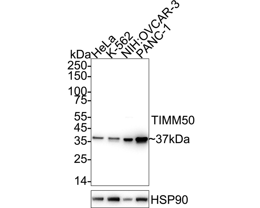
Western blot analysis of TIMM50 (Tim50) on different lysates with Rabbit anti-TIMM50 (Tim50) antibody at 1/5,000 dilution. Lane 1: HeLa cell lysate (20 µg/Lane), Lane 2: K-562 cell lysate (20 µg/Lane), Lane 3: NIH:OVCAR-3 cell lysate (20 µg/Lane), Lane 4: PANC-1 cell lysate (20 µg/Lane), Exposure time: 10 seconds; 4-20% SDS-PAGE gel. Proteins were transferred to a PVDF membrane and blocked with 5% NFDM/TBST for 1 hour at room temperature. The primary antibody at 1/5,000 dilution was used in primary antibody dilution at 4℃ overnight. Goat Anti-Rabbit IgG - HRP Secondary Antibody at 1/50,000 dilution was used for 1 hour at room temperature.IHC
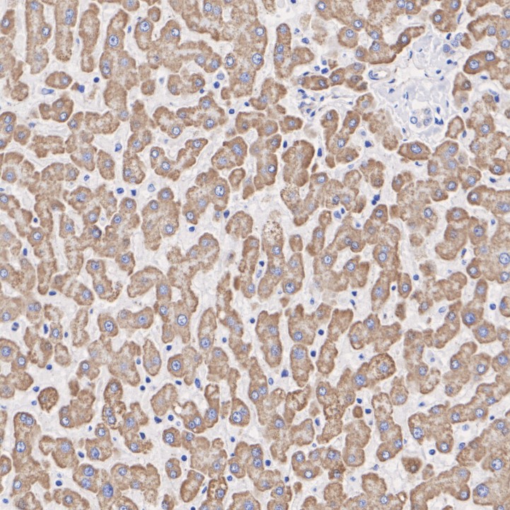
Immunohistochemical analysis of paraffin-embedded human liver tissue with Rabbit anti-TIMM50 (Tim50) antibody at 1/2,000 dilution. The section was pre-treated using heat mediated antigen retrieval with Tris-EDTA buffer (pH 9.0) for 20 minutes. The tissues were blocked in 1% BSA for 20 minutes at room temperature, washed with ddH2O and PBS, and then probed with the primary antibody at 1/2,000 dilution for 1 hour at room temperature. The detection was performed using an HRP conjugated compact polymer system. DAB was used as the chromogen. Tissues were counterstained with hematoxylin and mounted with DPX.IHC
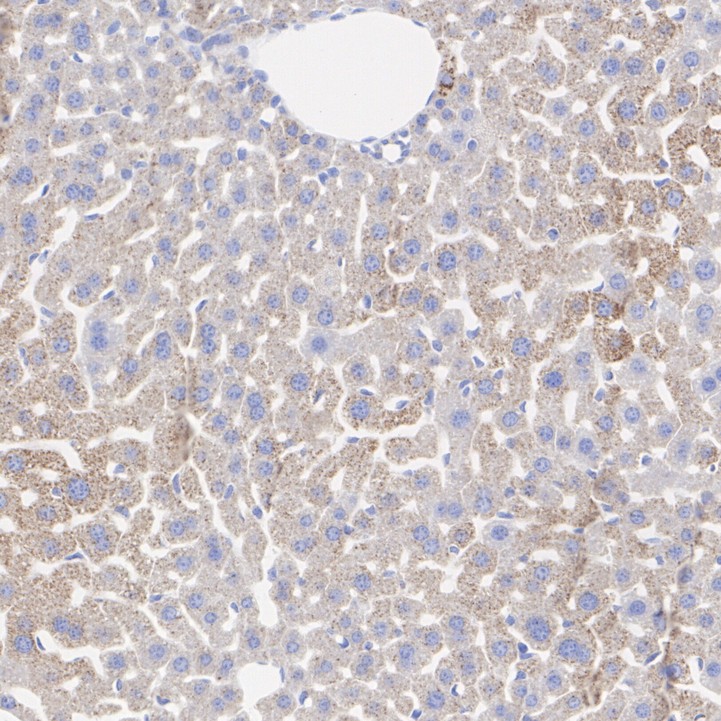
Immunohistochemical analysis of paraffin-embedded mouse liver tissue with Rabbit anti-TIMM50 (Tim50) antibody at 1/500 dilution. The section was pre-treated using heat mediated antigen retrieval with Tris-EDTA buffer (pH 9.0) for 20 minutes. The tissues were blocked in 1% BSA for 20 minutes at room temperature, washed with ddH2O and PBS, and then probed with the primary antibody at 1/500 dilution for 1 hour at room temperature. The detection was performed using an HRP conjugated compact polymer system. DAB was used as the chromogen. Tissues were counterstained with hematoxylin and mounted with DPX.IHC
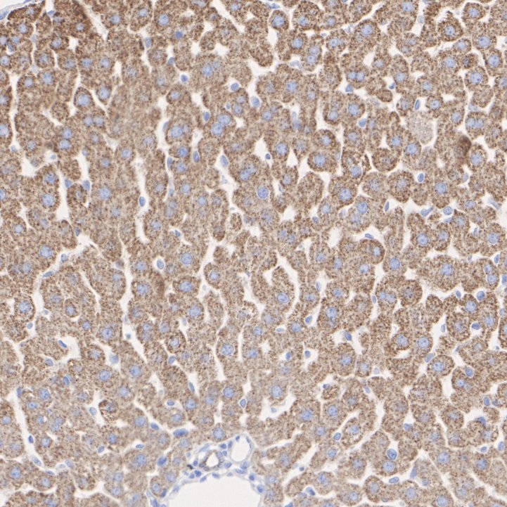
Immunohistochemical analysis of paraffin-embedded rat liver tissue with Rabbit anti-TIMM50 (Tim50) antibody at 1/500 dilution. The section was pre-treated using heat mediated antigen retrieval with Tris-EDTA buffer (pH 9.0) for 20 minutes. The tissues were blocked in 1% BSA for 20 minutes at room temperature, washed with ddH2O and PBS, and then probed with the primary antibody at 1/500 dilution for 1 hour at room temperature. The detection was performed using an HRP conjugated compact polymer system. DAB was used as the chromogen. Tissues were counterstained with hematoxylin and mounted with DPX.| Product Name | TIMM50 (Tim50) Recombinant Rabbit Monoclonal Antibody |
|---|---|
| Antibody Type | Primary Antibodies |
| Product description | Mitochondrial import inner membrane translocase subunit TIM50 is an enzyme that in humans is encoded by the TIMM50 gene. Tim50 is a subunit of the Tim23 translocase complex in the inner mitochondrial membrane. Mutations in TIMM50 can lead to epilepsy, severe intellectual disability, and 3-methylglutaconic aciduria. TIMM50 expression is increased in breast cancer cells and decreased in hypertrophic hearts. The TIMM50 gene is located on the q arm of chromosome 19 in position 13.2 and spans 13,373 base pairs. The gene produces a 39.6 kDa protein composed of 353 amino acids. This gene encodes a subunit of the TIM23 inner mitochondrial membrane translocase complex, which mediates the translocation of transit peptide-containing proteins across the mitochondrial inner membrane. The Tim50 protein functions as the receptor subunit that recognizes the mitochondrial targeting signal, or presequence, on protein cargo that is destined for the mitochondrial inner membrane and matrix. This gene in human cells results in the release of cytochrome c and apoptosis. This protein plays a role in maintaining the membrane permeability barrier. The intermembrane space domain of Tim50 induces the translocation pore of the TIM23 channel to close. |
| Immunogen | Recombinant protein within Human TIM50 aa 1-109 / 353. |
| Clonality | Monoclonal |
|---|---|
| Isotype | IgG |
| Host Species | Recombinant rabbit |
| Tested Applications | IHCWB |
| WB:1:5000 IHC:1:50-1:100 |
|
| Species Reactivity | HumanMouseRat |
| Concentration | 1mg/ |
| Purification | Protein A |
| Alternative Names | Homolog of yeast Tim50 antibody
Import inner membrane translocase subunit TIM 50 antibody
Import inner membrane translocase subunit TIM50 antibody Import inner membrane translocase subunit TIM50 mitochondrial antibody MGC102733 antibody Mitochondrial import inner membrane translocase subunit TIM50 antibody TIM 50 antibody TIM 50L antibody TIM50 antibody Tim50 like protein antibody TIM50_HUMAN antibody TIM50L antibody TIMM 50 antibody TIMM50 antibody translocase of inner mitochondrial membrane 50 homolog (S. cerevisiae) antibody Translocase of inner mitochondrial membrane 50 homolog (yeast) antibody Translocase of inner mitochondrial membrane 50 homolog antibody Translocase of inner mitochondrial membrane 50 yeast homolog of antibody |
|---|---|
| Molecular Weight(MW) | 40 kDa(Observed band size: 37 kDa) |
| Cellular Localization | Mitochondrion inner membrane, nucleus speckle. |
| SwissProt ID | Q3ZCQ8 |
|---|
WB
Western blot analysis of TIMM50 (Tim50) on different lysates with Rabbit anti-TIMM50 (Tim50) antibody at 1/5,000 dilution. Lane 1: HeLa cell lysate (20 µg/Lane), Lane 2: K-562 cell lysate (20 µg/Lane), Lane 3: NIH:OVCAR-3 cell lysate (20 µg/Lane), Lane 4: PANC-1 cell lysate (20 µg/Lane), Exposure time: 10 seconds; 4-20% SDS-PAGE gel. Proteins were transferred to a PVDF membrane and blocked with 5% NFDM/TBST for 1 hour at room temperature. The primary antibody at 1/5,000 dilution was used in primary antibody dilution at 4℃ overnight. Goat Anti-Rabbit IgG - HRP Secondary Antibody at 1/50,000 dilution was used for 1 hour at room temperature.
IHC
Immunohistochemical analysis of paraffin-embedded human liver tissue with Rabbit anti-TIMM50 (Tim50) antibody at 1/2,000 dilution. The section was pre-treated using heat mediated antigen retrieval with Tris-EDTA buffer (pH 9.0) for 20 minutes. The tissues were blocked in 1% BSA for 20 minutes at room temperature, washed with ddH2O and PBS, and then probed with the primary antibody at 1/2,000 dilution for 1 hour at room temperature. The detection was performed using an HRP conjugated compact polymer system. DAB was used as the chromogen. Tissues were counterstained with hematoxylin and mounted with DPX.
IHC
Immunohistochemical analysis of paraffin-embedded mouse liver tissue with Rabbit anti-TIMM50 (Tim50) antibody at 1/500 dilution. The section was pre-treated using heat mediated antigen retrieval with Tris-EDTA buffer (pH 9.0) for 20 minutes. The tissues were blocked in 1% BSA for 20 minutes at room temperature, washed with ddH2O and PBS, and then probed with the primary antibody at 1/500 dilution for 1 hour at room temperature. The detection was performed using an HRP conjugated compact polymer system. DAB was used as the chromogen. Tissues were counterstained with hematoxylin and mounted with DPX.
IHC
Immunohistochemical analysis of paraffin-embedded rat liver tissue with Rabbit anti-TIMM50 (Tim50) antibody at 1/500 dilution. The section was pre-treated using heat mediated antigen retrieval with Tris-EDTA buffer (pH 9.0) for 20 minutes. The tissues were blocked in 1% BSA for 20 minutes at room temperature, washed with ddH2O and PBS, and then probed with the primary antibody at 1/500 dilution for 1 hour at room temperature. The detection was performed using an HRP conjugated compact polymer system. DAB was used as the chromogen. Tissues were counterstained with hematoxylin and mounted with DPX.| Application Notes | WB:1:5000 IHC:1:50-1:100 |
|---|
| Form | Liquid |
|---|---|
| Storage Instructions | Store at +4℃ after thawing. Aliquot store at -20℃. Avoid repeated freeze / thaw cycles. |
| Storage Buffer | 1*TBS (pH7.4), 1%BSA, 50%Glycerol. Preservative: 0.05% Sodium Azide. |


