Application
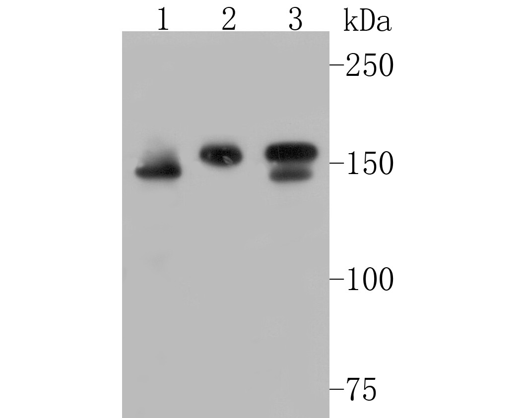
Fig1: Western blot analysis of WAPL on different lysates. Proteins were transferred to a PVDF membrane and blocked with 5% BSA in PBS for 1 hour at room temperature. The primary antibody (ET7110-25, 1/500) was used in 5% BSA at room temperature for 2 hours. Goat Anti-Rabbit IgG - HRP Secondary Antibody (HA1001) at 1:5,000 dilution was used for 1 hour at room temperature. Positive control: Lane 2: Hela cell lysate Lane 2: 293T cell lysate Lane 2: Jurkat cell lysateApplication
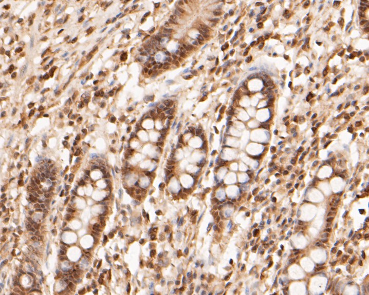
Fig2: Immunohistochemical analysis of paraffin-embedded human small intestine tissue using anti-WAPL antibody. The section was pre-treated using heat mediated antigen retrieval with sodium citrate buffer (pH 6.0) for 20 minutes. The tissues were blocked in 5% BSA for 30 minutes at room temperature, washed with ddH2O and PBS, and then probed with the primary antibody (ET7110-25, 1/100) for 30 minutes at room temperature. The detection was performed using an HRP conjugated compact polymer system. DAB was used as the chromogen. Tissues were counterstained with hematoxylin and mounted with DPX.Application
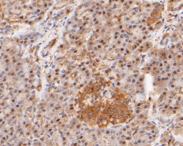
Fig3: Immunohistochemical analysis of paraffin-embedded human pancreas tissue using anti-WAPL antibody. The section was pre-treated using heat mediated antigen retrieval with sodium citrate buffer (pH 6.0) for 20 minutes. The tissues were blocked in 5% BSA for 30 minutes at room temperature, washed with ddH2O and PBS, and then probed with the primary antibody (ET7110-25, 1/100) for 30 minutes at room temperature. The detection was performed using an HRP conjugated compact polymer system. DAB was used as the chromogen. Tissues were counterstained with hematoxylin and mounted with DPX.Application
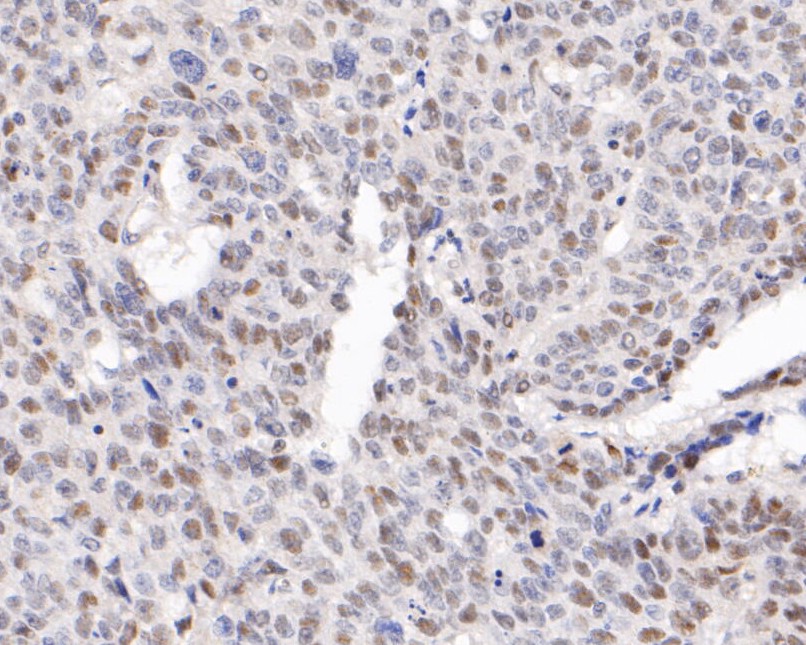
Fig4: Immunohistochemical analysis of paraffin-embedded human lung carcinoma tissue using anti-WAPL antibody. The section was pre-treated using heat mediated antigen retrieval with sodium citrate buffer (pH 6.0) for 20 minutes. The tissues were blocked in 5% BSA for 30 minutes at room temperature, washed with ddH2O and PBS, and then probed with the primary antibody (ET7110-25, 1/400) for 30 minutes at room temperature. The detection was performed using an HRP conjugated compact polymer system. DAB was used as the chromogen. Tissues were counterstained with hematoxylin and mounted with DPX.Application
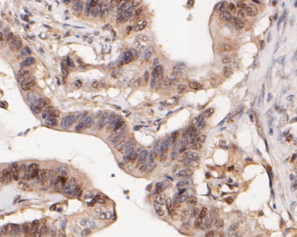
Fig5: Immunohistochemical analysis of paraffin-embedded human colon carcinoma tissue using anti-WAPL antibody. The section was pre-treated using heat mediated antigen retrieval with sodium citrate buffer (pH 6.0) for 20 minutes. The tissues were blocked in 5% BSA for 30 minutes at room temperature, washed with ddH2O and PBS, and then probed with the primary antibody (ET7110-25, 1/200) for 30 minutes at room temperature. The detection was performed using an HRP conjugated compact polymer system. DAB was used as the chromogen. Tissues were counterstained with hematoxylin and mounted with DPX.| Product Name | WAPL [JE51-75] |
|---|---|
| Antibody Type | Primary Antibodies |
| Product description | WAPL (wings apart-like), also known as WAPAL or FOE, is a 1,190 amino acid protein that contains one WAPL domain and is expressed as two alternatively spliced isoforms, one of which localizes to the nucleus. Expressed in an isoform-dependent manner in heart, skeletal muscle and uterine cervix tumor tissue, WAPL is involved in sister-chromatid adhesion and overall cell growth, specifically playing a role in the development and metastasis of cancerous tissue. The gene encoding WAPL maps to human chromosome 10, which houses over 1,200 genes and comprises nearly 4.5% of the human genome. Defects in some of the genes that map to chromosome 10 are associated with Charcot-Marie-Tooth disease, Jackson-Weiss syndrome, Usher syndrome, nonsyndromatic deafness, Wolman's syndrome, Cowden syndrome, multiple endocrine neoplasia type 2 and porphyria. |
| Immunogen | Synthetic peptide within N-terminal human WAPL |
| Clonality | Monoclonal |
|---|---|
| Isotype | IgG |
| Host Species | Recombinant rabbit |
| Tested Applications | WB,IHC-P |
| WB:1:500-1:2,000 IHC-P:1:50-1:500 |
|
| Species Reactivity | Human |
| Concentration | 1mg/mL |
| Purification | Unpurified |
| Alternative Names | FOE antibody
Friend of EBNA2 (Epstein Barr virus nuclear protein 2) antibody
Friend of EBNA2 protein antibody KIAA0261 antibody OTTHUMP00000020004 antibody WAPAL antibody WAPL antibody WAPL_HUMAN antibody Wings apart like homolog antibody Wings apart like homolog (Drosophila) antibody Wings apart-like protein homolog antibody |
|---|---|
| Molecular Weight(MW) | 133 kDa (Predicted band size) |
| Cellular Localization | Nucleus. |
| SwissProt ID | Q7Z5K2 |
|---|
Application
Fig1: Western blot analysis of WAPL on different lysates. Proteins were transferred to a PVDF membrane and blocked with 5% BSA in PBS for 1 hour at room temperature. The primary antibody (ET7110-25, 1/500) was used in 5% BSA at room temperature for 2 hours. Goat Anti-Rabbit IgG - HRP Secondary Antibody (HA1001) at 1:5,000 dilution was used for 1 hour at room temperature. Positive control: Lane 2: Hela cell lysate Lane 2: 293T cell lysate Lane 2: Jurkat cell lysate
Application
Fig2: Immunohistochemical analysis of paraffin-embedded human small intestine tissue using anti-WAPL antibody. The section was pre-treated using heat mediated antigen retrieval with sodium citrate buffer (pH 6.0) for 20 minutes. The tissues were blocked in 5% BSA for 30 minutes at room temperature, washed with ddH2O and PBS, and then probed with the primary antibody (ET7110-25, 1/100) for 30 minutes at room temperature. The detection was performed using an HRP conjugated compact polymer system. DAB was used as the chromogen. Tissues were counterstained with hematoxylin and mounted with DPX.
Application
Fig3: Immunohistochemical analysis of paraffin-embedded human pancreas tissue using anti-WAPL antibody. The section was pre-treated using heat mediated antigen retrieval with sodium citrate buffer (pH 6.0) for 20 minutes. The tissues were blocked in 5% BSA for 30 minutes at room temperature, washed with ddH2O and PBS, and then probed with the primary antibody (ET7110-25, 1/100) for 30 minutes at room temperature. The detection was performed using an HRP conjugated compact polymer system. DAB was used as the chromogen. Tissues were counterstained with hematoxylin and mounted with DPX.
Application
Fig4: Immunohistochemical analysis of paraffin-embedded human lung carcinoma tissue using anti-WAPL antibody. The section was pre-treated using heat mediated antigen retrieval with sodium citrate buffer (pH 6.0) for 20 minutes. The tissues were blocked in 5% BSA for 30 minutes at room temperature, washed with ddH2O and PBS, and then probed with the primary antibody (ET7110-25, 1/400) for 30 minutes at room temperature. The detection was performed using an HRP conjugated compact polymer system. DAB was used as the chromogen. Tissues were counterstained with hematoxylin and mounted with DPX.
Application
Fig5: Immunohistochemical analysis of paraffin-embedded human colon carcinoma tissue using anti-WAPL antibody. The section was pre-treated using heat mediated antigen retrieval with sodium citrate buffer (pH 6.0) for 20 minutes. The tissues were blocked in 5% BSA for 30 minutes at room temperature, washed with ddH2O and PBS, and then probed with the primary antibody (ET7110-25, 1/200) for 30 minutes at room temperature. The detection was performed using an HRP conjugated compact polymer system. DAB was used as the chromogen. Tissues were counterstained with hematoxylin and mounted with DPX.| Positive Control | Hela cell lysates, 293T cell lysates, Jurkat cell lysates, human small intestine tissue, human pancreas tissue, human lung carcinoma tissue, human colon carcinoma tissue. |
|---|---|
| Application Notes | WB:1:500-1:2,000 IHC-P:1:50-1:500 |
| Form | Liquid |
|---|---|
| Storage Instructions | Store at +4℃ after thawing. Aliquot store at -20℃. Avoid repeated freeze / thaw cycles. |
| Storage Buffer | 1*TBS (pH7.4), 1%BSA, 50%Glycerol. Preservative: 0.05% Sodium Azide. |


