Application
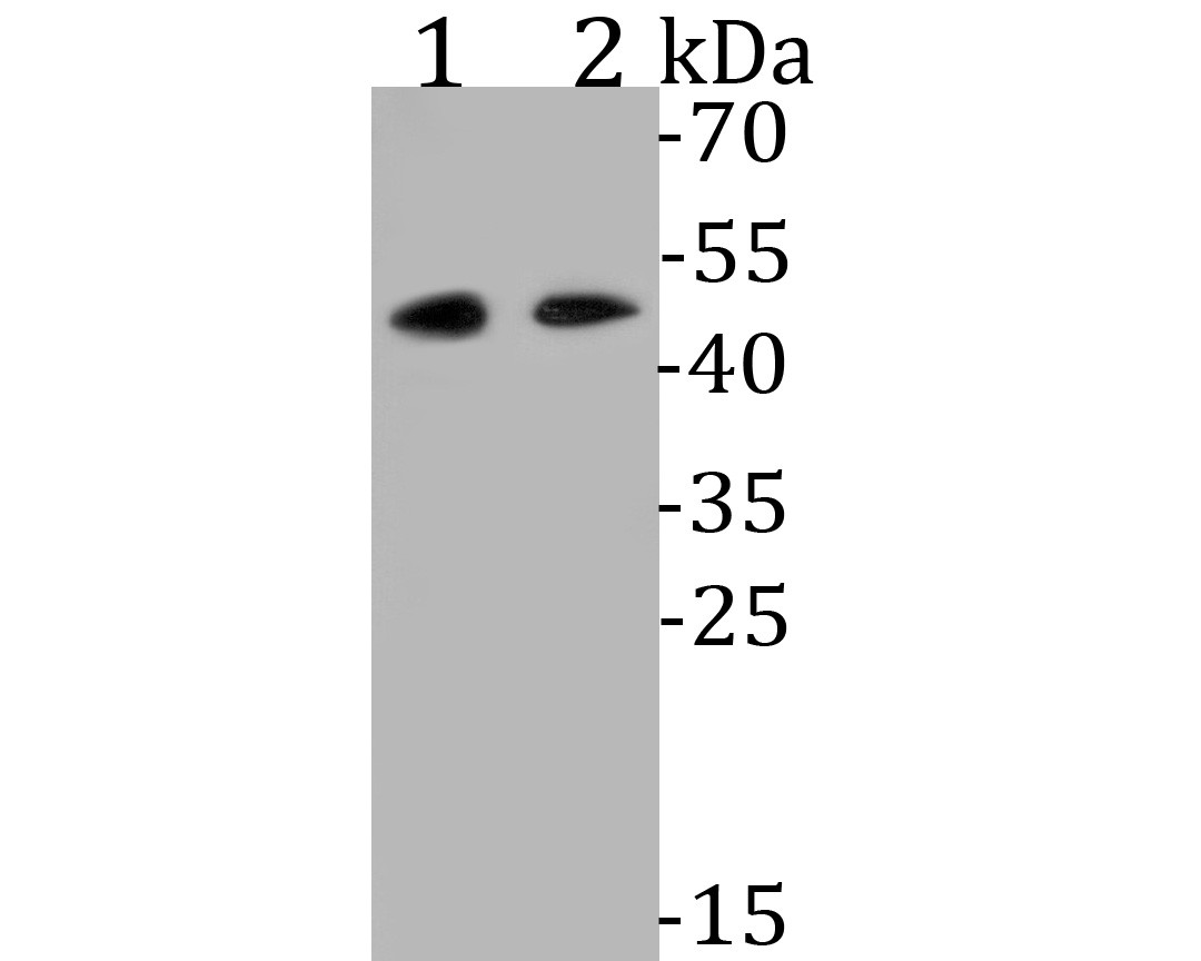
Fig1: Western blot analysis of WNT5B on different lysates. Proteins were transferred to a PVDF membrane and blocked with 5% BSA in PBS for 1 hour at room temperature. The primary antibody (ET7110-34, 1/500) was used in 5% BSA at room temperature for 2 hours. Goat Anti-Rabbit IgG - HRP Secondary Antibody (HA1001) at 1:5,000 dilution was used for 1 hour at room temperature. Positive control: Lane 1: Hela cell lysate Lane 2: HepG2 cell lysateApplication
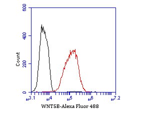
Fig2: Flow cytometric analysis of WNT5B was done on LOVO cells. The cells were fixed, permeabilized and stained with the primary antibody (ET7110-34, 1/50) (red). After incubation of the primary antibody at room temperature for an hour, the cells were stained with a Alexa Fluor 488-conjugated Goat anti-Rabbit IgG Secondary antibody at 1/1000 dilution for 30 minutes.Unlabelled sample was used as a control (cells without incubation with primary antibody; black).| Product Name | WNT5B [JE53-67] |
|---|---|
| Antibody Type | Primary Antibodies |
| Antigen Alias | MGC2648 antibody Protein Wnt-5b antibody Wingless type MMTV integration site family, member 5B antibody WNT 5B antibody WNT 5B protein antibody Wnt family member 5B antibody Wnt5b antibody WNT5B protein antibody WNT5B_HUMAN antibody |
| Product description | Protein Wnt-5b is a protein that in humans is encoded by the WNT5B gene. The WNT gene family consists of structurally related genes which encode secreted signaling proteins. These proteins have been implicated in oncogenesis and in several developmental processes, including regulation of cell fate and patterning during embryogenesis. This gene is a member of the WNT gene family. It encodes a protein which shows 94% and 80% amino acid identity to the mouse Wnt5b protein and the human WNT5A protein, respectively. Alternative splicing of this gene generates 2 transcript variants. |
| Immunogen | Synthetic peptide within C terminal human Wnt5b. |
| Clonality | Monoclonal |
|---|---|
| Isotype | IgG |
| Host Species | Recombinant rabbit |
| Tested Applications | WBFC |
| WB:1:500-1:2,000: FC:1:50-1:100: |
|
| Species Reactivity | Human |
| Concentration | 1mg/ml |
| Alternative Names | MGC2648 antibody
Protein Wnt-5b antibody
Wingless type MMTV integration site family member 5B antibody WNT 5B antibody WNT 5B protein antibody Wnt family member 5B antibody Wnt5b antibody WNT5B protein antibody WNT5B_HUMAN antibody |
|---|---|
| Molecular Weight(MW) | Predicted band size 40 kDa. |
| Cellular Localization | Extracellular matrix. |
| SwissProt ID | Q9H1J7 |
|---|
Application
Fig1: Western blot analysis of WNT5B on different lysates. Proteins were transferred to a PVDF membrane and blocked with 5% BSA in PBS for 1 hour at room temperature. The primary antibody (ET7110-34, 1/500) was used in 5% BSA at room temperature for 2 hours. Goat Anti-Rabbit IgG - HRP Secondary Antibody (HA1001) at 1:5,000 dilution was used for 1 hour at room temperature. Positive control: Lane 1: Hela cell lysate Lane 2: HepG2 cell lysate
Application
Fig2: Flow cytometric analysis of WNT5B was done on LOVO cells. The cells were fixed, permeabilized and stained with the primary antibody (ET7110-34, 1/50) (red). After incubation of the primary antibody at room temperature for an hour, the cells were stained with a Alexa Fluor 488-conjugated Goat anti-Rabbit IgG Secondary antibody at 1/1000 dilution for 30 minutes.Unlabelled sample was used as a control (cells without incubation with primary antibody; black).| Positive Control | Hela cell lysate, HepG2 cell lysate, LOVO. |
|---|---|
| Application Notes | WB:1:500-1:2,000: FC:1:50-1:100: |
| Form | Liquid |
|---|---|
| Storage Instructions | Store at +4℃ after thawing. Aliquot store at -20℃. Avoid repeated freeze / thaw cycles. |
| Storage Buffer | 1*TBS (pH7.4), 1%BSA, 50%Glycerol. Preservative: 0.05% Sodium Azide. |


