Application
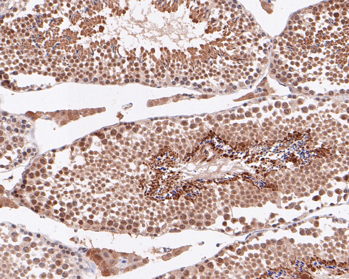
Fig3: Immunohistochemical analysis of paraffin-embedded mouse testis tissue using anti-ABI2 antibody. The section was pre-treated using heat mediated antigen retrieval with sodium citrate buffer (pH 6.0) for 20 minutes. The tissues were blocked in 5% BSA for 30 minutes at room temperature, washed with ddH2O and PBS, and then probed with the primary antibody (ET7110-42, 1/50) for 30 minutes at room temperature. The detection was performed using an HRP conjugated compact polymer system. DAB was used as the chromogen. Tissues were counterstained with hematoxylin and mounted with DPX.Application
Fig3: Immunohistochemical analysis of paraffin-embedded mouse testis tissue using anti-ABI2 antibody. The section was pre-treated using heat mediated antigen retrieval with sodium citrate buffer (pH 6.0) for 20 minutes. The tissues were blocked in 5% BSA for 30 minutes at room temperature, washed with ddH2O and PBS, and then probed with the primary antibody (ET7110-42, 1/50) for 30 minutes at room temperature. The detection was performed using an HRP conjugated compact polymer system. DAB was used as the chromogen. Tissues were counterstained with hematoxylin and mounted with DPX.Application
Fig3: Immunohistochemical analysis of paraffin-embedded mouse testis tissue using anti-ABI2 antibody. The section was pre-treated using heat mediated antigen retrieval with sodium citrate buffer (pH 6.0) for 20 minutes. The tissues were blocked in 5% BSA for 30 minutes at room temperature, washed with ddH2O and PBS, and then probed with the primary antibody (ET7110-42, 1/50) for 30 minutes at room temperature. The detection was performed using an HRP conjugated compact polymer system. DAB was used as the chromogen. Tissues were counterstained with hematoxylin and mounted with DPX.| Product Name | ABI2 [JE53-89] |
|---|---|
| Antibody Type | Primary Antibodies |
| Antigen Alias | Abelson interactor 2 antibody ABI 2 antibody Abi-2 antibody ABI2 antibody ABI2_HUMAN antibody ABI2B antibody Abl binding protein 3 antibody Abl interacting protein 1 (SH3 containing protein) antibody Abl interactor 2 antibody Abl interactor protein 2b antibody Abl-binding protein 3 antibody AblBP3 antibody AIP 1 antibody Arg binding protein 1 antibody Arg protein tyrosine kinase binding protein antibody Arg-binding protein 1 antibody ArgBP1 antibody ARGBPIA antibody ArgBPIB antibody OTTHUMP00000163783 antibody OTTHUMP00000163784 antibody OTTHUMP00000206182 antibody OTTHUMP00000206183 antibody OTTHUMP00000206184 antibody OTTHUMP00000206185 antibody SSH3BP2 antibody |
| Product description | Regulator of actin cytoskeleton dynamics underlying cell motility and adhesion. Functions as a component of the WAVE complex, which activates actin nucleating machinery Arp2/3 to drive lamellipodia formation. Acts as regulator and substrate of nonreceptor tyrosine kinases ABL1 and ABL2 involved in processes linked to cell growth and differentiation. Positively regulates ABL1-mediated phosphorylation of ENAH, which is required for proper polymerization of nucleated actin filaments at the leading edge. Contributes to the regulation of actin assembly at the tips of neuron projections. In particular, controls dendritic spine morphogenesis and may promote dendritic spine specification toward large mushroom-type spines known as repositories of memory in the brain (By similarity). In hippocampal neurons, may mediate actin-dependent BDNF-NTRK2 early endocytic trafficking that triggers dendrite outgrowth (By similarity). Participates in ocular lens morphogenesis, likely by regulating lamellipodia-driven adherens junction formation at the epithelial cell-secondary lens fiber interface (By similarity). Also required for nascent adherens junction assembly in epithelial cells. |
| Immunogen | Synthetic peptide within N terminal human ABI2. |
| Clonality | Monoclonal |
|---|---|
| Isotype | IgG |
| Host Species | Recombinant rabbit |
| Tested Applications | WBIHC-P |
| WB:1:500-1:2,000: IHC-P:1:50-1:200: |
|
| Species Reactivity | HumanMouse |
| Concentration | 1mg/ml |
| Alternative Names | Abelson interactor 2 antibody
ABI 2 antibody
Abi-2 antibody ABI2 antibody ABI2_HUMAN antibody ABI2B antibody Abl binding protein 3 antibody Abl interacting protein 1 (SH3 containing protein) antibody Abl interactor 2 antibody Abl interactor protein 2b antibody Abl-binding protein 3 antibody AblBP3 antibody AIP 1 antibody Arg binding protein 1 antibody Arg protein tyrosine kinase binding protein antibody Arg-binding protein 1 antibody ArgBP1 antibody ARGBPIA antibody ArgBPIB antibody OTTHUMP00000163783 antibody OTTHUMP00000163784 antibody OTTHUMP00000206182 antibody OTTHUMP00000206183 antibody OTTHUMP00000206184 antibody OTTHUMP00000206185 antibody SSH3BP2 antibody |
|---|---|
| Molecular Weight(MW) | 56 kDa |
| Cellular Localization | Nucleus, Cytoplasm. |
Application
Fig3: Immunohistochemical analysis of paraffin-embedded mouse testis tissue using anti-ABI2 antibody. The section was pre-treated using heat mediated antigen retrieval with sodium citrate buffer (pH 6.0) for 20 minutes. The tissues were blocked in 5% BSA for 30 minutes at room temperature, washed with ddH2O and PBS, and then probed with the primary antibody (ET7110-42, 1/50) for 30 minutes at room temperature. The detection was performed using an HRP conjugated compact polymer system. DAB was used as the chromogen. Tissues were counterstained with hematoxylin and mounted with DPX.
Application
Fig3: Immunohistochemical analysis of paraffin-embedded mouse testis tissue using anti-ABI2 antibody. The section was pre-treated using heat mediated antigen retrieval with sodium citrate buffer (pH 6.0) for 20 minutes. The tissues were blocked in 5% BSA for 30 minutes at room temperature, washed with ddH2O and PBS, and then probed with the primary antibody (ET7110-42, 1/50) for 30 minutes at room temperature. The detection was performed using an HRP conjugated compact polymer system. DAB was used as the chromogen. Tissues were counterstained with hematoxylin and mounted with DPX.
Application
Fig3: Immunohistochemical analysis of paraffin-embedded mouse testis tissue using anti-ABI2 antibody. The section was pre-treated using heat mediated antigen retrieval with sodium citrate buffer (pH 6.0) for 20 minutes. The tissues were blocked in 5% BSA for 30 minutes at room temperature, washed with ddH2O and PBS, and then probed with the primary antibody (ET7110-42, 1/50) for 30 minutes at room temperature. The detection was performed using an HRP conjugated compact polymer system. DAB was used as the chromogen. Tissues were counterstained with hematoxylin and mounted with DPX.| Positive Control | MCF-7 cell lysates, human lung tissue, mouse testis tissue. |
|---|---|
| Application Notes | WB:1:500-1:2,000: IHC-P:1:50-1:200: |
| Form | Liquid |
|---|---|
| Storage Instructions | Store at +4℃ after thawing. Aliquot store at -20℃. Avoid repeated freeze / thaw cycles. |
| Storage Buffer | 1*TBS (pH7.4), 1%BSA, 50%Glycerol. Preservative: 0.05% Sodium Azide. |


