Application
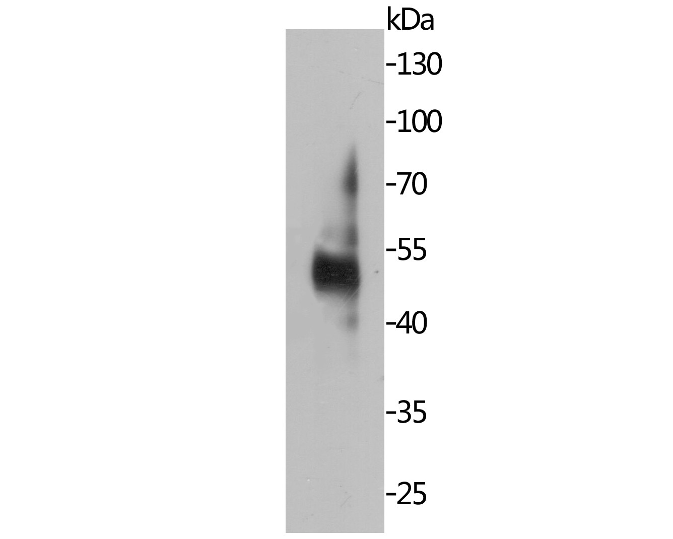
Fig1: Western blot analysis of MUM1 on SKBR3 lysates. Proteins were transferred to a PVDF membrane and blocked with 5% BSA in PBS for 1 hour at room temperature. The primary antibody (, 1/500) was used in 5% BSA at room temperature for 2 hours. Goat Anti-Rabbit IgG - HRP Secondary Antibody (HA1001) at 1:5,000 dilution was used for 1 hour at room temperature.Application
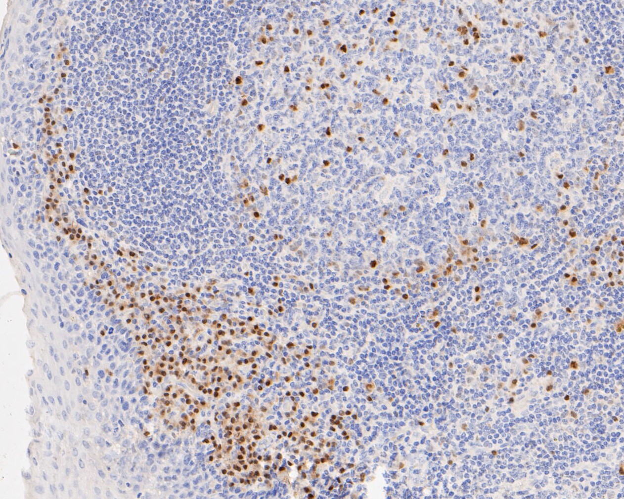
Fig2: Immunohistochemical analysis of paraffin-embedded human tonsil tissue using anti-MUM1 antibody. The section was pre-treated using heat mediated antigen retrieval with sodium citrate buffer (pH 6.0) for 20 minutes. The tissues were blocked in 5% BSA for 30 minutes at room temperature, washed with ddH2O and PBS, and then probed with the primary antibody (ET7110-44, 1/50) for 30 minutes at room temperature. The detection was performed using an HRP conjugated compact polymer system. DAB was used as the chromogen. Tissues were counterstained with hematoxylin and mounted with DPX.Application
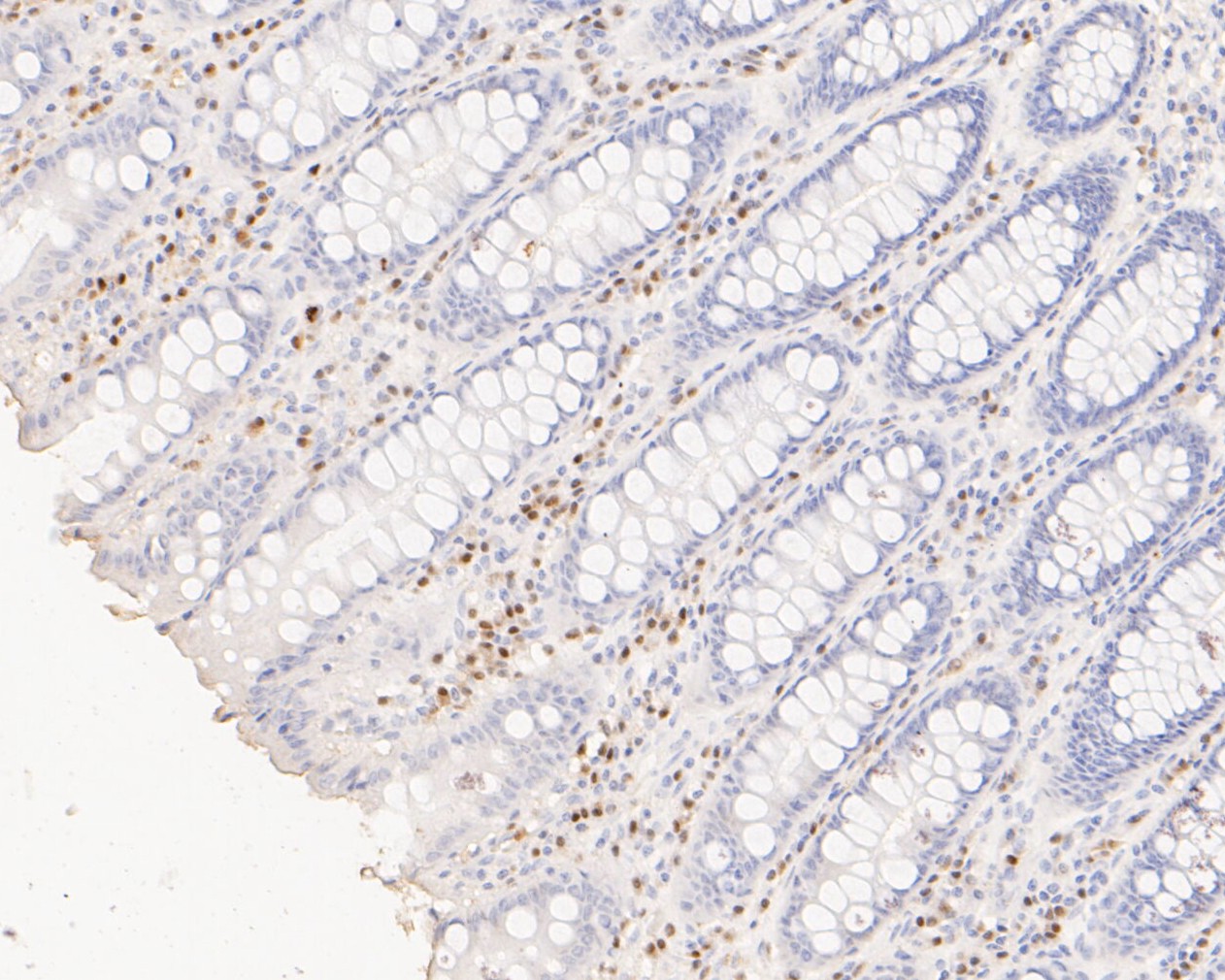
Fig3: Immunohistochemical analysis of paraffin-embedded human colon tissue using anti-MUM1 antibody. The section was pre-treated using heat mediated antigen retrieval with sodium citrate buffer (pH 6.0) for 20 minutes. The tissues were blocked in 5% BSA for 30 minutes at room temperature, washed with ddH2O and PBS, and then probed with the primary antibody (ET7110-44, 1/50) for 30 minutes at room temperature. The detection was performed using an HRP conjugated compact polymer system. DAB was used as the chromogen. Tissues were counterstained with hematoxylin and mounted with DPX.| Product Name | MUM1 [JE52-60] |
|---|---|
| Antibody Type | Primary Antibodies |
| Antigen Alias | "Interferon regulatory factor 4 antibody IRF 4 antibody IRF-4 antibody Irf4 antibody IRF4_HUMAN antibody LSIRF antibody Lymphocyte specific interferon regulatory factor antibody Lymphocyte specific IRF antibody Lymphocyte-specific interferon regulatory factor antibody Multiple myeloma oncogene 1 antibody MUM 1 antibody MUM1 antibody NF EM5 antibody NF-EM5 antibody NFEM5 antibody PU.1 interaction partner antibody Sfpi1/PU.1 interaction partner antibody Transcriptional activator PIP antibody" |
| Product description | Interferon regulatory factor 4 also known as MUM1 is a protein that in humans is encoded by the IRF4 gene, located at 6p25-p23. In melanocytic cells the IRF4 gene may be regulated by MITF. IRF4 is a transcription factor that has been implicated in acute leukemia. This gene is strongly associated with pigmentation: sensitivity of skin to sun exposure, freckles, blue eyes, and brown hair color. A variant has been implicated in greying of hair. The World Health Organization (2016) provisionally defined large B-cell lymphoma with IRF4 rearrangement as a rare indolent large B-cell lymphoma of children and adolescents. This indolent lymphoma mimics, and must be distinguished from, pediatric-type follicular lymphoma. The hallmark of large B-cell lymphoma with IRF4 rearrangement is the overexpression of the IRF4 gene by the disease's malignant cells. Transcriptional activator. Binds to the interferon-stimulated response element (ISRE) of the MHC class I promoter. Binds the immunoglobulin lambda light chain enhancer, together with PU.1. Probably plays a role in ISRE-targeted signal transduction mechanisms specific to lymphoid cells. Involved in CD8+ dendritic cell differentiation by forming a complex with the BATF-JUNB heterodimer in immune cells, leading to recognition of AICE sequence (5'-TGAnTCA/GAAA-3'), an immune-specific regulatory element, followed by cooperative binding of BATF and IRF4 and activation of genes. |
| Immunogen | Synthetic peptide within N terminal human MUM1. |
| Clonality | Monoclonal |
|---|---|
| Isotype | IgG |
| Host Species | Recombinant rabbit |
| Tested Applications | WBIHC |
| WB:1:500-1:2,000: IHC:1:50-1:200: |
|
| Species Reactivity | Human |
| Concentration | 1mg/ml |
| Alternative Names | "Interferon regulatory factor 4 antibody
IRF 4 antibody
IRF-4 antibody Irf4 antibody IRF4_HUMAN antibody LSIRF antibody Lymphocyte specific interferon regulatory factor antibody Lymphocyte specific IRF antibody Lymphocyte-specific interferon regulatory factor antibody Multiple myeloma oncogene 1 antibody MUM 1 antibody MUM1 antibody NF EM5 antibody NF-EM5 antibody NFEM5 antibody PU.1 interaction partner antibody Sfpi1/PU.1 interaction partner antibody Transcriptional activator PIP antibody" |
|---|---|
| Molecular Weight(MW) | 52 kDa |
| Cellular Localization | Nucleus. |
| SwissProt ID | Q15306 |
|---|
Application
Fig1: Western blot analysis of MUM1 on SKBR3 lysates. Proteins were transferred to a PVDF membrane and blocked with 5% BSA in PBS for 1 hour at room temperature. The primary antibody (, 1/500) was used in 5% BSA at room temperature for 2 hours. Goat Anti-Rabbit IgG - HRP Secondary Antibody (HA1001) at 1:5,000 dilution was used for 1 hour at room temperature.
Application
Fig2: Immunohistochemical analysis of paraffin-embedded human tonsil tissue using anti-MUM1 antibody. The section was pre-treated using heat mediated antigen retrieval with sodium citrate buffer (pH 6.0) for 20 minutes. The tissues were blocked in 5% BSA for 30 minutes at room temperature, washed with ddH2O and PBS, and then probed with the primary antibody (ET7110-44, 1/50) for 30 minutes at room temperature. The detection was performed using an HRP conjugated compact polymer system. DAB was used as the chromogen. Tissues were counterstained with hematoxylin and mounted with DPX.
Application
Fig3: Immunohistochemical analysis of paraffin-embedded human colon tissue using anti-MUM1 antibody. The section was pre-treated using heat mediated antigen retrieval with sodium citrate buffer (pH 6.0) for 20 minutes. The tissues were blocked in 5% BSA for 30 minutes at room temperature, washed with ddH2O and PBS, and then probed with the primary antibody (ET7110-44, 1/50) for 30 minutes at room temperature. The detection was performed using an HRP conjugated compact polymer system. DAB was used as the chromogen. Tissues were counterstained with hematoxylin and mounted with DPX.| Positive Control | Human tonsil tissue, human colon tissue. |
|---|---|
| Application Notes | WB:1:500-1:2,000: IHC:1:50-1:200: |
| Form | Liquid |
|---|---|
| Storage Instructions | Store at +4℃ after thawing. Aliquot store at -20℃. Avoid repeated freeze / thaw cycles. |
| Storage Buffer | 1*TBS (pH7.4), 1%BSA, 50%Glycerol. Preservative: 0.05% Sodium Azide. |


