Application
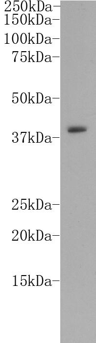
Fig1: Western blot analysis on K562 cell lysates using anti-CD14 rabbit polyclonal antibody.Application
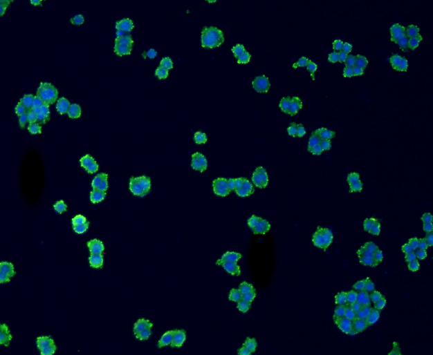
Fig2: ICC staining CD14 in SW480 cells (green). The nuclear counter stain is DAPI (blue). Cells were fixed in paraformaldehyde, permeabilised with 0.25% Triton X100/PBS.Application
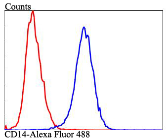
Fig3: Flow cytometric analysis of Raji cells with CD14 antibody at 1/100 dilution (blue) compared with an unlabelled control (cells without incubation with primary antibody; red). Alexa Fluor 488-conjugated goat anti-rabbit IgG was used as the secondary antibody.| Product Name | CD14 |
|---|---|
| Antibody Type | Primary Antibodies |
| Antigen Alias | CD 14 antibody CD_antigen=CD14 antibody CD14 antibody CD14 antigen antibody CD14 molecule antibody CD14_HUMAN antibody LPS-R antibody Mo2 antibody Monocyte differentiation antigen CD14 antibody Monocyte differentiation antigen CD14 urinary form antibody Monocyte differentiation antigen CD14, membrane-bound form antibody Myeloid cell specific leucine rich glycoprotein antibody Myeloid cell-specific leucine-rich glycoprotein antibody |
| Product description | CD14 is a glycophosphatidylinositol-anchored protein expressed on the surfaces of monocytes and plymorphonuclear leukocytes. It cooperates with MD-2 and TLR4 to mediate the innate immune response to bacterial lipopolysaccharide (LPS). CD14 acts via MyD88, TIRAP and TRAF6, leading to NF-kappa-B activation, cytokine secretion and the inflammatory response and up-regulates cell surface molecules, including adhesion molecules. |
| Immunogen | Synthetic peptide (KLH-coupled) within human CD14 150-210 aa. |
| Clonality | Polyclonal |
|---|---|
| Isotype | IgG |
| Host Species | Rabbit |
| Tested Applications | WBICCFC |
| WB:1:1,000 ICC:1:50 FC:1:50 |
|
| Species Reactivity | HumanMouse |
| Concentration | 1mg/ml |
| Alternative Names | CD 14 antibody
CD_antigen=CD14 antibody
CD14 antibody
CD14 antigen antibody
CD14 molecule antibody
CD14_HUMAN antibody
LPS-R antibody
Mo2 antibody
Monocyte differentiation antigen CD14 antibody
Monocyte differentiation antigen CD14 urinary form antibody
Monocyte differentiation antigen CD14 membrane-bound form antibody Myeloid cell specific leucine rich glycoprotein antibody Myeloid cell-specific leucine-rich glycoprotein antibody |
|---|---|
| Molecular Weight(MW) | 39kDa |
| Cellular Localization | Cell membrane |
Application
Fig1: Western blot analysis on K562 cell lysates using anti-CD14 rabbit polyclonal antibody.
Application
Fig2: ICC staining CD14 in SW480 cells (green). The nuclear counter stain is DAPI (blue). Cells were fixed in paraformaldehyde, permeabilised with 0.25% Triton X100/PBS.
Application
Fig3: Flow cytometric analysis of Raji cells with CD14 antibody at 1/100 dilution (blue) compared with an unlabelled control (cells without incubation with primary antibody; red). Alexa Fluor 488-conjugated goat anti-rabbit IgG was used as the secondary antibody.| Positive Control | K562, SW480, Raji |
|---|---|
| Application Notes | WB:1:1,000 ICC:1:50 FC:1:50 |
| Form | Liquid |
|---|---|
| Storage Instructions | Store at +4℃ after thawing. Aliquot store at -20℃. Avoid repeated freeze / thaw cycles. |
| Storage Buffer | 1*TBS (pH7.4), 1%BSA, 40%Glycerol. Preservative: 0.05% Sodium Azide. |


