Application
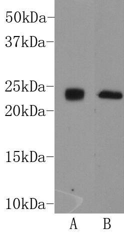
Fig1: Western blot analysis on mouse brain (A) and human brain (B) cell lysates using anti-THY-1 rabbit polyclonal antibody.Application
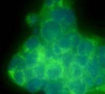
Fig2:ICC image of THY-1 antibody stained NCCIT cells. The secondary antibody (green) was goat anti-rabbit IgG (H+L) FITC conjugated. DAPI was used to stain the cell nuclei (blue).| Product Name | THY-1 |
|---|---|
| Antibody Type | Primary Antibodies |
| Antigen Alias | CD7 antibody CD90 antibody CD90 antigen antibody CDw90 antibody FLJ33325 antibody MGC128895 antibody T25 antibody Theta antigen antibody Thy 1 antibody Thy 1 cell surface antigen antibody Thy 1 membrane glycoprotein antibody Thy 1 T cell antigen antibody Thy 1.2 antibody Thy-1 antigen antibody Thy-1 membrane glycoprotein antibody Thy1 antibody Thy1 antigen antibody Thy1 T cell antigen antibody Thy1.1 antibody Thy1.2 antibody THY1_HUMAN antibody Thymus cell antigen 1, theta antibody |
| Product description | Thy-1 or CD90 is a 25–37 kDa heavily N-glycosylated, glycophosphatidylinositol (GPI) anchored conserved cell surface protein with a single V-like immunoglobulin domain, originally discovered as a thymocyte antigen. It is highly expressed on the surface of adult neurons and is thought to play a role in modulating adhesive and migratory events, such as neurite extension. Thy-1 can be used as a marker for a variety of stem cells and for the axonal processes of mature neurons. |
| Immunogen | peptide |
| Clonality | Polyclonal |
|---|---|
| Isotype | IgG |
| Host Species | Rabbit |
| Tested Applications | WBICC |
| WB:1:500-1:1,000 ICC:1:50-1:100 |
|
| Species Reactivity | HumanMouse |
| Concentration | 1mg/ml |
| Alternative Names | CD7 antibody
CD90 antibody
CD90 antigen antibody
CDw90 antibody
FLJ33325 antibody
MGC128895 antibody
T25 antibody
Theta antigen antibody
Thy 1 antibody
Thy 1 cell surface antigen antibody
Thy 1 membrane glycoprotein antibody
Thy 1 T cell antigen antibody
Thy 1.2 antibody
Thy-1 antigen antibody
Thy-1 membrane glycoprotein antibody
Thy1 antibody
Thy1 antigen antibody
Thy1 T cell antigen antibody
Thy1.1 antibody
Thy1.2 antibody
THY1_HUMAN antibody
Thymus cell antigen 1 theta antibody |
|---|---|
| Molecular Weight(MW) | 22kDa |
| Cellular Localization | Cell membrane |
| SwissProt ID | P04216 |
|---|
Application
Fig1: Western blot analysis on mouse brain (A) and human brain (B) cell lysates using anti-THY-1 rabbit polyclonal antibody.
Application
Fig2:ICC image of THY-1 antibody stained NCCIT cells. The secondary antibody (green) was goat anti-rabbit IgG (H+L) FITC conjugated. DAPI was used to stain the cell nuclei (blue).| Positive Control | Human brain, mouse brain, NCCIT |
|---|---|
| Application Notes | WB:1:500-1:1,000 ICC:1:50-1:100 |
| Form | Liquid |
|---|---|
| Storage Instructions | Store at +4℃ after thawing. Aliquot store at -20℃ or -80℃. Avoid repeated freeze / thaw cycles. |
| Storage Buffer | 1*TBS (pH7.4), 1%BSA, 40%Glycerol. Preservative: 0.05% Sodium Azide. |


