WB
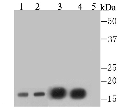
Western blot analysis of Histone H3 (di methyl K4) on different lysates. Proteins were transferred to a PVDF membrane and blocked with 5% BSA in PBS for 1 hour at room temperature. The primary antibody was used at a 1:1,000 dilution in 5% BSA at room temperature for 2 hours. Goat Anti-Rabbit IgG - HRP Secondary Antibody (HA1001) at 1:5,000 dilution was used for 1 hour at room temperature. Positive control: Lane 1: Human liver tissue lysate, untreated, Lane 2: Histone lysate, purified from 293T, Lane 3: F9 cell lysate, untreated, Lane 4: F9 cell lysate, treated with Histone H3 peptide-unmodifed, Lane 5: F9 cell lysate, treated with Histone H3 peptide-di-methyl K4.IHC
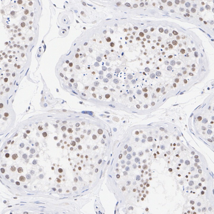
Immunohistochemical analysis of paraffin-embedded human testis tissue with Rabbit anti-Histone H3 (di methyl K4) antibody at 1/3,000 dilution. The section was pre-treated using heat mediated antigen retrieval with sodium citrate buffer (pH 6.0) (high pressure) for 2 minutes. The tissues were blocked in 1% BSA for 20 minutes at room temperature, washed with ddH2O and PBS, and then probed with the primary antibody at 1/3,000 dilution for 1 hour at room temperature. The detection was performed using an HRP conjugated compact polymer system. DAB was used as the chromogen. Tissues were counterstained with hematoxylin and mounted with DPX.IHC
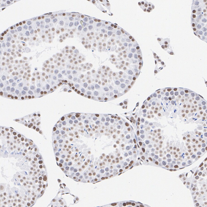
Immunohistochemical analysis of paraffin-embedded mouse testis tissue with Rabbit anti-Histone H3 (di methyl K4) antibody at 1/3,000 dilution. The section was pre-treated using heat mediated antigen retrieval with sodium citrate buffer (pH 6.0) (high pressure) for 2 minutes. The tissues were blocked in 1% BSA for 20 minutes at room temperature, washed with ddH2O and PBS, and then probed with the primary antibody at 1/3,000 dilution for 1 hour at room temperature. The detection was performed using an HRP conjugated compact polymer system. DAB was used as the chromogen. Tissues were counterstained with hematoxylin and mounted with DPX.IHC
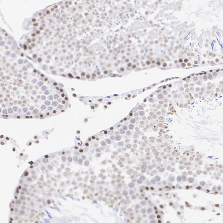
Immunohistochemical analysis of paraffin-embedded rat testis tissue with Rabbit anti-Histone H3 (di methyl K4) antibody at 1/3,000 dilution. The section was pre-treated using heat mediated antigen retrieval with sodium citrate buffer (pH 6.0) (high pressure) for 2 minutes. The tissues were blocked in 1% BSA for 20 minutes at room temperature, washed with ddH2O and PBS, and then probed with the primary antibody at 1/3,000 dilution for 1 hour at room temperature. The detection was performed using an HRP conjugated compact polymer system. DAB was used as the chromogen. Tissues were counterstained with hematoxylin and mounted with DPX.ICC/IF
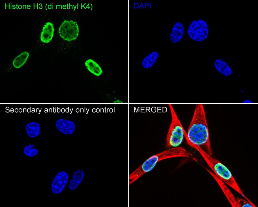
Immunocytochemistry analysis of MCF7 cells labeling Histone H3 (di methyl K4) with Rabbit anti-Histone H3 (di methyl K4) antibody at 1/5,000 dilution. Cells were fixed in 4% paraformaldehyde for 20 minutes at room temperature, permeabilized with 0.1% Triton X-100 in PBS for 5 minutes at room temperature, then blocked with 1% BSA in 10% negative goat serum for 1 hour at room temperature. Cells were then incubated with Rabbit anti-Histone H3 (di methyl K4) antibody at 1/5,000 dilution in 1% BSA in PBST overnight at 4 ℃. Goat Anti-Rabbit IgG H&L (488) was used as the secondary antibody at 1/1,000 dilution. PBS instead of the primary antibody was used as the secondary antibody only control. Nuclear DNA was labelled in blue with DAPI. Beta tubulin (red) was stained at 1/100 dilution overnight at +4℃. Goat Anti-Mouse IgG H&L (594) was used as the secondary antibody at 1/1,000 dilution.FC
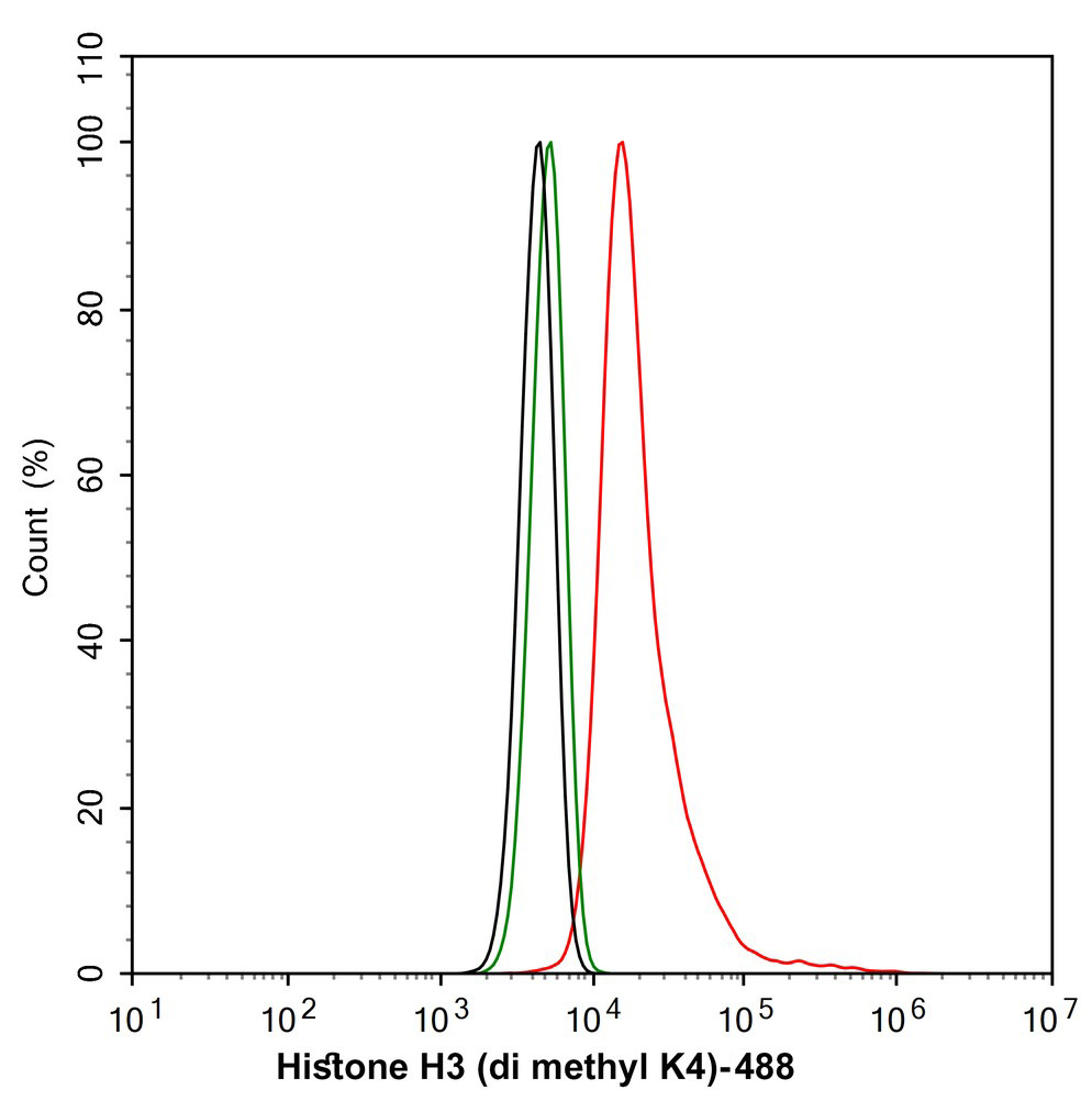
Flow cytometric analysis of NIH/3T3 cells labeling Histone H3 (di methyl K4). Cells were fixed and permeabilized. Then stained with the primary antibody (1/1,000) (red) compared with Rabbit IgG Isotype Control (green). After incubation of the primary antibody at +4℃ for an hour, the cells were stained with a 488 conjugate-Goat anti-Rabbit IgG Secondary antibody at 1/1,000 dilution for 30 minutes at +4℃. Unlabelled sample was used as a control (cells without incubation with primary antibody; black).DB
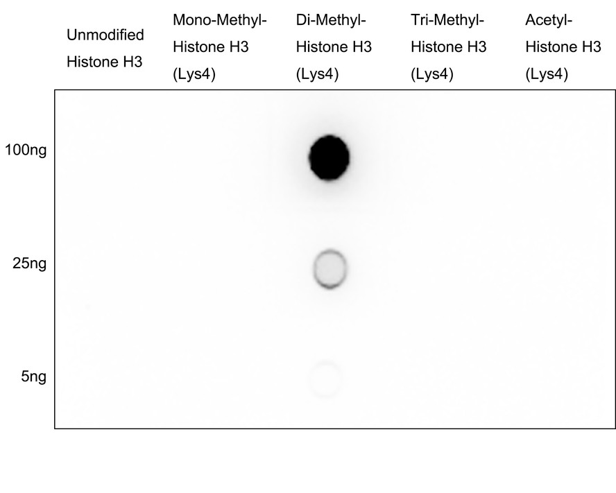
Dot blot analysis of Histone H3 (di methyl K4) on different proteins with Rabbit anti-Histone H3 (di methyl K4) antibody at 1/1,000 dilution. Goat Anti-Rabbit IgG - HRP Secondary Antibody at 1/50,000 dilution for 1 hour at room temperature. Lane 1: Unmodified Histone H3 (negative), Lane 2: Mono-Methyl-Histone H3 (Lys4) (negative), Lane 3: Di-Methyl-Histone H3 (Lys4) (positive), Lane 4: Tri-Methyl-Histone H3 (Lys4) (negative), Lane 5: Acetyl-Histone H3 (Lys4) (negative), Proteins loading: 100ng, 25ng, 5ng; Blocking and dilution buffer: 5% NFDM/TBST; Exposure time: 1 minute 30 seconds.| Product Name | Histone H3 (di methyl K4) Rabbit Polyclonal Antibody |
|---|---|
| Antibody Type | Primary Antibodies |
| Product description | The nucleosome, made up of DNA wound around eight core histone proteins (two each of H2A, H2B, H3, and H4), is the primary building block of chromatin. Histones thereby play a central role in transcription regulation, DNA repair, DNA replication and chromosomal stability. DNA accessibility is regulated via a complex set of post-translational modifications of histones, also called histone code, and nucleosome remodeling. Methylation at Lys-5 (H3K4me), Lys-37 (H3K36me) and Lys-80 (H3K79me) are linked to gene activation. Methylation at Lys-5 (H3K4me) facilitates subsequent acetylation of H3 and H4. Methylation at Lys-80 (H3K79me) is associated with DNA double-strand break (DSB) responses and is a specific target for TP53BP1. Methylation at Lys-10 (H3K9me) and Lys-28 (H3K27me) are linked to gene repression. Methylation at Lys-10 (H3K9me) is a specific target for HP1 proteins (CBX1, CBX3 and CBX5) and prevents subsequent phosphorylation at Ser-11 (H3S10ph) and acetylation of H3 and H4. Methylation at Lys-5 (H3K4me) and Lys-80 (H3K79me) require preliminary monoubiquitination of H2B at 'Lys-120'. Methylation at Lys-10 (H3K9me) and Lys-28 (H3K27me) are enriched in inactive X chromosome chromatin. |
| Immunogen | Synthetic peptide of the N terminal residues of Human Di-mehtyl-Histone H3(Lys4). |
| Clonality | Polyclonal |
|---|---|
| Isotype | IgG |
| Host Species | Rabbit |
| Tested Applications | ChIPFCICC/IFIHCWB |
| WB:1:1000 IHC:1:50-1:200 ICC/IF:1:500-1:5000 FC:1:1000 DB:1:1000 |
|
| Species Reactivity | HumanMouseRat |
| Concentration | 1mg/ml |
| Purification | Protein A |
| Alternative Names | Histone H3/a Histone H3/b Histone H3/c Histone H3/d Histone H3/f Histone H3/h Histone H3/i Histone H3/j Histone H3/k Histone H3/l H3K4me2 |
|---|---|
| Molecular Weight(MW) | 15kDa |
| Cellular Localization | Nucleus |
| SwissProt ID | P68431 |
|---|
WB
Western blot analysis of Histone H3 (di methyl K4) on different lysates. Proteins were transferred to a PVDF membrane and blocked with 5% BSA in PBS for 1 hour at room temperature. The primary antibody was used at a 1:1,000 dilution in 5% BSA at room temperature for 2 hours. Goat Anti-Rabbit IgG - HRP Secondary Antibody (HA1001) at 1:5,000 dilution was used for 1 hour at room temperature. Positive control: Lane 1: Human liver tissue lysate, untreated, Lane 2: Histone lysate, purified from 293T, Lane 3: F9 cell lysate, untreated, Lane 4: F9 cell lysate, treated with Histone H3 peptide-unmodifed, Lane 5: F9 cell lysate, treated with Histone H3 peptide-di-methyl K4.
IHC
Immunohistochemical analysis of paraffin-embedded human testis tissue with Rabbit anti-Histone H3 (di methyl K4) antibody at 1/3,000 dilution. The section was pre-treated using heat mediated antigen retrieval with sodium citrate buffer (pH 6.0) (high pressure) for 2 minutes. The tissues were blocked in 1% BSA for 20 minutes at room temperature, washed with ddH2O and PBS, and then probed with the primary antibody at 1/3,000 dilution for 1 hour at room temperature. The detection was performed using an HRP conjugated compact polymer system. DAB was used as the chromogen. Tissues were counterstained with hematoxylin and mounted with DPX.
IHC
Immunohistochemical analysis of paraffin-embedded mouse testis tissue with Rabbit anti-Histone H3 (di methyl K4) antibody at 1/3,000 dilution. The section was pre-treated using heat mediated antigen retrieval with sodium citrate buffer (pH 6.0) (high pressure) for 2 minutes. The tissues were blocked in 1% BSA for 20 minutes at room temperature, washed with ddH2O and PBS, and then probed with the primary antibody at 1/3,000 dilution for 1 hour at room temperature. The detection was performed using an HRP conjugated compact polymer system. DAB was used as the chromogen. Tissues were counterstained with hematoxylin and mounted with DPX.
IHC
Immunohistochemical analysis of paraffin-embedded rat testis tissue with Rabbit anti-Histone H3 (di methyl K4) antibody at 1/3,000 dilution. The section was pre-treated using heat mediated antigen retrieval with sodium citrate buffer (pH 6.0) (high pressure) for 2 minutes. The tissues were blocked in 1% BSA for 20 minutes at room temperature, washed with ddH2O and PBS, and then probed with the primary antibody at 1/3,000 dilution for 1 hour at room temperature. The detection was performed using an HRP conjugated compact polymer system. DAB was used as the chromogen. Tissues were counterstained with hematoxylin and mounted with DPX.
ICC/IF
Immunocytochemistry analysis of MCF7 cells labeling Histone H3 (di methyl K4) with Rabbit anti-Histone H3 (di methyl K4) antibody at 1/5,000 dilution. Cells were fixed in 4% paraformaldehyde for 20 minutes at room temperature, permeabilized with 0.1% Triton X-100 in PBS for 5 minutes at room temperature, then blocked with 1% BSA in 10% negative goat serum for 1 hour at room temperature. Cells were then incubated with Rabbit anti-Histone H3 (di methyl K4) antibody at 1/5,000 dilution in 1% BSA in PBST overnight at 4 ℃. Goat Anti-Rabbit IgG H&L (488) was used as the secondary antibody at 1/1,000 dilution. PBS instead of the primary antibody was used as the secondary antibody only control. Nuclear DNA was labelled in blue with DAPI. Beta tubulin (red) was stained at 1/100 dilution overnight at +4℃. Goat Anti-Mouse IgG H&L (594) was used as the secondary antibody at 1/1,000 dilution.
FC
Flow cytometric analysis of NIH/3T3 cells labeling Histone H3 (di methyl K4). Cells were fixed and permeabilized. Then stained with the primary antibody (1/1,000) (red) compared with Rabbit IgG Isotype Control (green). After incubation of the primary antibody at +4℃ for an hour, the cells were stained with a 488 conjugate-Goat anti-Rabbit IgG Secondary antibody at 1/1,000 dilution for 30 minutes at +4℃. Unlabelled sample was used as a control (cells without incubation with primary antibody; black).
DB
Dot blot analysis of Histone H3 (di methyl K4) on different proteins with Rabbit anti-Histone H3 (di methyl K4) antibody at 1/1,000 dilution. Goat Anti-Rabbit IgG - HRP Secondary Antibody at 1/50,000 dilution for 1 hour at room temperature. Lane 1: Unmodified Histone H3 (negative), Lane 2: Mono-Methyl-Histone H3 (Lys4) (negative), Lane 3: Di-Methyl-Histone H3 (Lys4) (positive), Lane 4: Tri-Methyl-Histone H3 (Lys4) (negative), Lane 5: Acetyl-Histone H3 (Lys4) (negative), Proteins loading: 100ng, 25ng, 5ng; Blocking and dilution buffer: 5% NFDM/TBST; Exposure time: 1 minute 30 seconds.| Application Notes | WB:1:1000 IHC:1:50-1:200 ICC/IF:1:500-1:5000 FC:1:1000 DB:1:1000 |
|---|
| Form | Liquid |
|---|---|
| Storage Instructions | Store at +4℃ after thawing. Aliquot store at -20℃ or -80℃. Avoid repeated freeze / thaw cycles. |
| Storage Buffer | 1*PBS (pH7.4), 0.2% BSA, 40% Glycerol. Preservative: 0.05% Sodium Azide. |


