Application
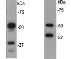
Fig1: Western blot analysis on F9 (A) and NCCIT (B) cell lysates using CD79a rabbit polyclonal antibody.Application
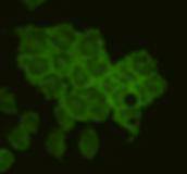
Fig2:ICC image of CD79a antibody stained F9 cells. The secondary antibody (green) was goat anti-rabbit IgG (H+L) FITC conjugated.Application
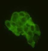
Fig3:ICC image of CD79a antibody stained NCCIT cells. The secondary antibody (green) was goat anti-rabbit IgG (H+L) FITC conjugated.| Product Name | CD79a |
|---|---|
| Antibody Type | Primary Antibodies |
| Antigen Alias | B lymphocyte-specific MB1 protein antibody B-cell antigen receptor complex-associated protein alpha chain antibody CD 79a antibody CD79a antibody CD79a antigen (immunoglobulin-associated alpha) antibody CD79A antigen antibody CD79a molecule, immunoglobulin-associated alpha antibody CD79A_HUMAN antibody Ig alpha antibody Ig-alpha antibody IGA antibody IgM-alpha antibody Immunoglobulin-associated alpha antibody Ly54 antibody MB-1 membrane glycoprotein antibody MB1 antibody Membrane-bound immunoglobulin-associated protein antibody Surface IgM-associated protein antibody |
| Product description | The mb-1 gene codes for a phosphoprotein, designated CD79a, that, together with the related CD79b protein, forms a dimer associated with membrane-bound immunoglobulin in B-cells. The CD79a/CD79b dimer is closely associated with the B-cell antigen receptor, in a similar manner to the association of CD3 with the T-cell receptor, and enables the cell to respond to the presence of antigens on its surface. The CD79a protein is present on the surface of B-cells throughout their life cycle, and is absent on all other healthy cells, making it a highly reliable marker for B-cells in immunohistochemistry |
| Immunogen | peptide |
| Clonality | Polyclonal |
|---|---|
| Isotype | IgG |
| Host Species | Rabbit |
| Tested Applications | WBICC |
| WB:1 ICC:1:50-1:100 |
|
| Species Reactivity | HumanMouse |
| Concentration | 1mg/ml |
| Alternative Names | B lymphocyte-specific MB1 protein antibody
B-cell antigen receptor complex-associated protein alpha chain antibody
CD 79a antibody
CD79a antibody
CD79a antigen (immunoglobulin-associated alpha) antibody
CD79A antigen antibody
CD79a molecule immunoglobulin-associated alpha antibody CD79A_HUMAN antibody Ig alpha antibody Ig-alpha antibody IGA antibody IgM-alpha antibody Immunoglobulin-associated alpha antibody Ly54 antibody MB-1 membrane glycoprotein antibody MB1 antibody Membrane-bound immunoglobulin-associated protein antibody Surface IgM-associated protein antibody |
|---|---|
| Molecular Weight(MW) | 40-50kDa |
| Cellular Localization | Cell membrane |
| SwissProt ID | P11911 |
|---|
Application
Fig1: Western blot analysis on F9 (A) and NCCIT (B) cell lysates using CD79a rabbit polyclonal antibody.
Application
Fig2:ICC image of CD79a antibody stained F9 cells. The secondary antibody (green) was goat anti-rabbit IgG (H+L) FITC conjugated.
Application
Fig3:ICC image of CD79a antibody stained NCCIT cells. The secondary antibody (green) was goat anti-rabbit IgG (H+L) FITC conjugated.| Positive Control | F9, NCCIT |
|---|---|
| Application Notes | WB:1 ICC:1:50-1:100 |
| Form | Liquid |
|---|---|
| Storage Instructions | Store at +4℃ after thawing. Aliquot store at -20℃ or -80℃. Avoid repeated freeze / thaw cycles. |
| Storage Buffer | 1*TBS (pH7.4), 1%BSA, 40%Glycerol. Preservative: 0.05% Sodium Azide. |


