Application
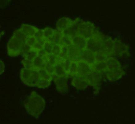
Fig1:ICC image of Rem2 antibody stained F9 cells. The secondary antibody (green) was goat anti-rabbit IgG (H+L) FITC conjugated.Application
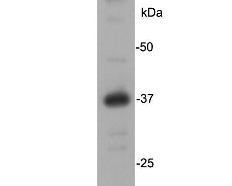
Fig2: Western blot analysis on NCCIT cell lysates using anti-Rem2 rabbit polyclonal antibody.Application
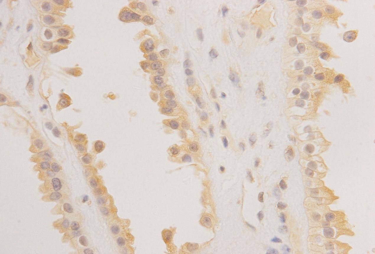
Fig3: IHC image of Rem2 antibody on human spleen sections.| Product Name | Rem2 |
|---|---|
| Antibody Type | Primary Antibodies |
| Antigen Alias | FLJ38964 antibody GTP binding protein REM 2 antibody Rad and Gem like GTP binding protein 2 antibody Rad and Gem related 2 antibody Rad and gem related GTP binding protein 2 antibody RAS (RAD and GEM) like GTP binding 2 antibody REM 2 antibody |
| Product description | REM2, a Ras-like GTPase, may be involved in increasing endothelial cell proliferation and the induction of angiogenesis. REM2 belongs to the small GTPase superfamily, RGK family. It binds GTP saturably and exhibits a low intrinsic rate of GTP hydrolysis |
| Immunogen | peptide |
| Clonality | Polyclonal |
|---|---|
| Isotype | IgG |
| Host Species | Rabbit |
| Tested Applications | WBICCIHC |
| WB:1:1,000 ICC:1:20 IHC:1:200-1:400 |
|
| Species Reactivity | HumanMouse |
| Concentration | 1mg/ml |
| Alternative Names | FLJ38964 antibody GTP binding protein REM 2 antibody Rad and Gem like GTP binding protein 2 antibody Rad and Gem related 2 antibody Rad and gem related GTP binding protein 2 antibody RAS (RAD and GEM) like GTP binding 2 antibody REM 2 antibody |
|---|---|
| Molecular Weight(MW) | 37kDa |
| Cellular Localization | Cell membrane, Cytoplasm |
| SwissProt ID | Q8IYK8 |
|---|
Application
Fig1:ICC image of Rem2 antibody stained F9 cells. The secondary antibody (green) was goat anti-rabbit IgG (H+L) FITC conjugated.
Application
Fig2: Western blot analysis on NCCIT cell lysates using anti-Rem2 rabbit polyclonal antibody.
Application
Fig3: IHC image of Rem2 antibody on human spleen sections.| Positive Control | F9, NCCIT, Human spleen |
|---|---|
| Application Notes | WB:1:1,000 ICC:1:20 IHC:1:200-1:400 |
| Form | Liquid |
|---|---|
| Storage Instructions | Store at +4℃ after thawing. Aliquot store at -20℃ or -80℃. Avoid repeated freeze / thaw cycles. |
| Storage Buffer | 1*TBS (pH7.4), 1%BSA, 40%Glycerol. Preservative: 0.05% Sodium Azide. |


