Application
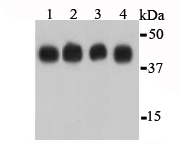
Fig1: Western blot analysis of β-actin on different lysates using anti-β-actin antibody at 1/1,000 dilution.Positive control:Lane 1: NIH/3T3Lane 2: HelaLane 3: PC12Lane 4: ZebrafishApplication
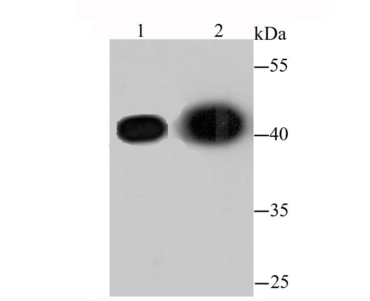
Fig2: Western blot analysis of β-actin on different lysates using anti-β-actin antibody at 1/1,000 dilution.Positive control:Lane 1: hybrid fish (crucian-carp) brain tissueLane 2: hybrid fish (crucian-carp) kidney tissue| Product Name | β-Actin |
|---|---|
| Antibody Type | Primary Antibodies |
| Antigen Alias | A26C1A antibody A26C1B antibody ACTB antibody ACTB_HUMAN antibody Actin beta antibody Actin cytoplasmic 1 antibody Actin, cytoplasmic 1, N-terminally processed antibody Actx antibody b actin antibody Beta cytoskeletal actin antibody Beta-actin antibody BRWS1 antibody E430023M04Rik antibody MGC128179 antibody PS1TP5 binding protein 1 antibody PS1TP5BP1 antibody |
| Product description | Actins are highly conserved proteins involved in cell motility, structure and integrity. Actin has been found to be expressed in at least six isomeric forms. It is expressed in heart and skeletal striated muscle tissue, and in certain smooth muscle tissues, regulating contractile potentials for these cells. It is also expressed in the cytoplasm of non-muscle cells, functioning to control cell structure and motility. Beta actin is usually used as a loading control, for among others, the integrity of cells, protein degradation, in Western Blotting. |
| Immunogen | peptide |
| Clonality | Polyclonal |
|---|---|
| Isotype | IgG |
| Host Species | Rabbit |
| Tested Applications | WBICCIHCFC |
| WB:1:1,000-5,000 |
|
| Species Reactivity | HumanMouseRatZebra FishOther |
| Concentration | 1mg/ml |
| Alternative Names | A26C1A antibody
A26C1B antibody
ACTB antibody
ACTB_HUMAN antibody
Actin beta antibody
Actin cytoplasmic 1 antibody
Actin cytoplasmic 1 N-terminally processed antibody Actx antibody b actin antibody Beta cytoskeletal actin antibody Beta-actin antibody BRWS1 antibody E430023M04Rik antibody MGC128179 antibody PS1TP5 binding protein 1 antibody PS1TP5BP1 antibody |
|---|---|
| Molecular Weight(MW) | 42kDa |
| Cellular Localization | Cytoskeleton |
| SwissProt ID | P60709 |
|---|
Application
Fig1: Western blot analysis of β-actin on different lysates using anti-β-actin antibody at 1/1,000 dilution.Positive control:Lane 1: NIH/3T3Lane 2: HelaLane 3: PC12Lane 4: Zebrafish
Application
Fig2: Western blot analysis of β-actin on different lysates using anti-β-actin antibody at 1/1,000 dilution.Positive control:Lane 1: hybrid fish (crucian-carp) brain tissueLane 2: hybrid fish (crucian-carp) kidney tissue| Positive Control | Hela, NIH/3T3, PC12, Zebrafish |
|---|---|
| Application Notes | WB:1:1,000-5,000 |
| Form | Liquid |
|---|---|
| Storage Instructions | Store at +4℃ after thawing. Aliquot store at -20℃ or -80℃. Avoid repeated freeze / thaw cycles. |
| Storage Buffer | 1*TBS (pH7.4), 1%BSA, 40%Glycerol. Preservative: 0.05% Sodium Azide. |


