WB
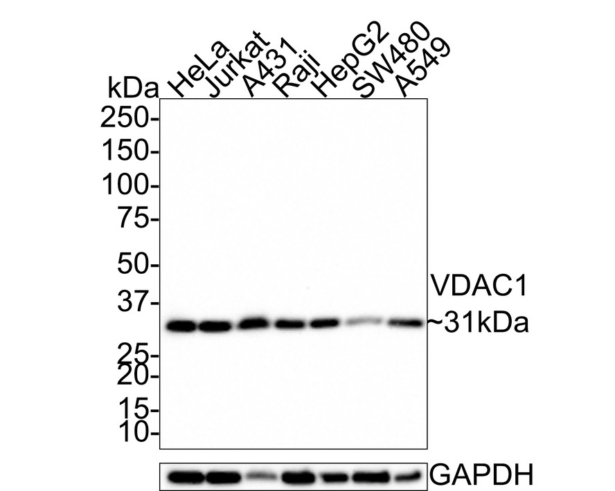
Western blot analysis of VDAC1 on different lysates with Rabbit anti-VDAC1 antibody at 1/1,000 dilution. Lane 1: HeLa cell lysate, Lane 2: Jurkat cell lysate, Lane 3: A431 cell lysate, Lane 4: Raji cell lysate, Lane 5: HepG2 cell lysate, Lane 6: SW480 cell lysate, Lane 7: A549 cell lysate, Lysates/proteins at 20 µg/Lane. Exposure time: 1 minute 30 seconds; 4-20% SDS-PAGE gel. Proteins were transferred to a PVDF membrane and blocked with 5% NFDM/TBST for 1 hour at room temperature. The primary antibody at 1/1,000 dilution was used in 5% NFDM/TBST at room temperature for 2 hours. Goat Anti-Rabbit IgG - HRP Secondary Antibody at 1:100,000 dilution was used for 1 hour at room temperature.IHC
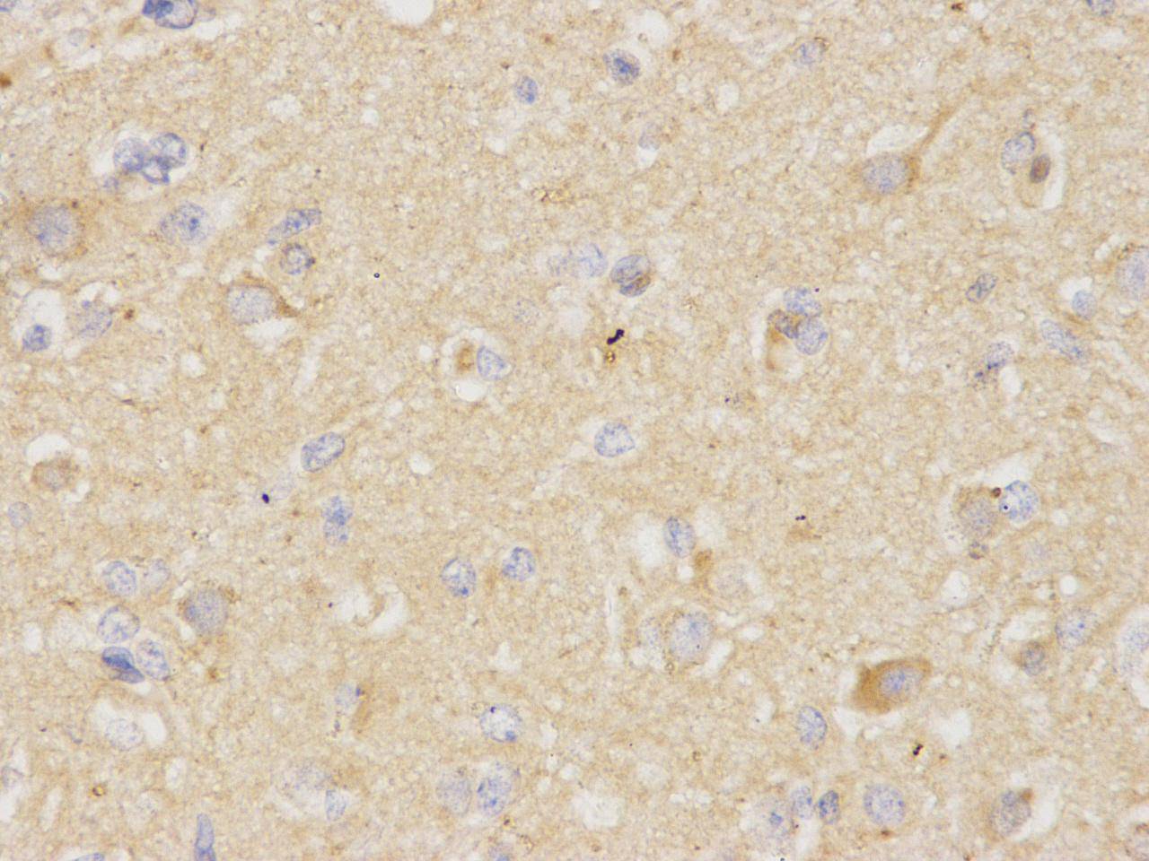
Immunohistochemical analysis of paraffin-embedded human brain tissue using anti-VDAC1 rabbit polyclonal antibody.ICC/IF
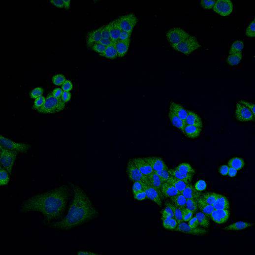
ICC staining VDAC1 in Hela cells (green). Cells were fixed in paraformaldehyde, permeabilised with 0.25% Triton X100/PBS and counterstained with DAPI in order to highlight the nucleus (blue).| Product Name | VDAC1 Rabbit Polyclonal Antibody |
|---|---|
| Antibody Type | Primary Antibodies |
| Product description | Voltage-dependent anion-selective channel (VDAC1) (also referred to as porin, isoform 1) is a small protein, originally discovered in the outer membrane of mitochondria where it constitutes the major pore-forming protein. The porin protein VDAC1 allows to the outer-most membrane of the mitochondrion free permeability to low molecular-weight solutes. VDAC1 has been shown to co-immunoprecipitate with the anti-apoptotic protein Bcl-2 and suggested to be involved in forming the mitochondrial pore which releases cytochrome c during apoptosis. |
| Immunogen | Synthetic peptide within human VDAC1 aa 188-231. |
| Clonality | Polyclonal |
|---|---|
| Isotype | IgG |
| Host Species | Rabbit |
| Tested Applications | ICC/IFIHCWB |
| WB:1:1000 IHC:1:200 ICC/IF:1:200 |
|
| Species Reactivity | HumanZebrafish |
| Concentration | 1mg/ml |
| Purification | Affinity purified |
| Alternative Names | hVDAC1 antibody Outer mitochondrial membrane protein porin 1 antibody Plasmalemmal porin antibody Porin 31HL antibody Porin 31HM antibody Porin antibody VDAC 1 antibody VDAC antibody VDAC-1 antibody Vdac1 antibody VDAC1_HUMAN antibody Voltage Dependent Anion Channel 1 antibody Voltage dependent anion selective channel protein 1 antibody Voltage-dependent anion-selective channel protein 1 antibody |
|---|---|
| Molecular Weight(MW) | 31kDa |
| Cellular Localization | Mitochondrial membrane, cell membrane |
| SwissProt ID | P21796 |
|---|
WB
Western blot analysis of VDAC1 on different lysates with Rabbit anti-VDAC1 antibody at 1/1,000 dilution. Lane 1: HeLa cell lysate, Lane 2: Jurkat cell lysate, Lane 3: A431 cell lysate, Lane 4: Raji cell lysate, Lane 5: HepG2 cell lysate, Lane 6: SW480 cell lysate, Lane 7: A549 cell lysate, Lysates/proteins at 20 µg/Lane. Exposure time: 1 minute 30 seconds; 4-20% SDS-PAGE gel. Proteins were transferred to a PVDF membrane and blocked with 5% NFDM/TBST for 1 hour at room temperature. The primary antibody at 1/1,000 dilution was used in 5% NFDM/TBST at room temperature for 2 hours. Goat Anti-Rabbit IgG - HRP Secondary Antibody at 1:100,000 dilution was used for 1 hour at room temperature.
IHC
Immunohistochemical analysis of paraffin-embedded human brain tissue using anti-VDAC1 rabbit polyclonal antibody.
ICC/IF
ICC staining VDAC1 in Hela cells (green). Cells were fixed in paraformaldehyde, permeabilised with 0.25% Triton X100/PBS and counterstained with DAPI in order to highlight the nucleus (blue).| Positive Control | A431, human brain, Hela, Raji, HepG2, SW480 |
|---|---|
| Application Notes | WB:1:1000 IHC:1:200 ICC/IF:1:200 |
| Form | Liquid |
|---|---|
| Storage Instructions | Store at +4℃ after thawing. Aliquot store at -20℃ or -80℃. Avoid repeated freeze / thaw cycles. |
| Storage Buffer | 1*TBS (pH7.4), 0.5%BSA, 40%Glycerol. Preservative: 0.05% Sodium Azide. |


