Application
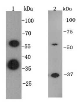
Fig1: Western blot analysis on A549 (1) and F9 (2) cell lysates using anti-ESRRB rabbit polyclonal antibodies.Application
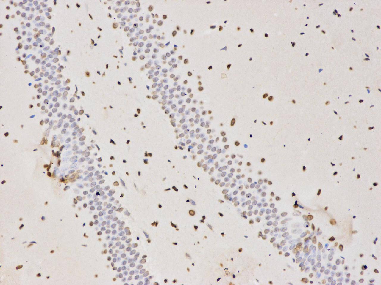
Fig2:Immunohistochemical analysis of paraffin-embedded rat brain tissue using anti- ESRRB rabbit polyclonal antibody.Application
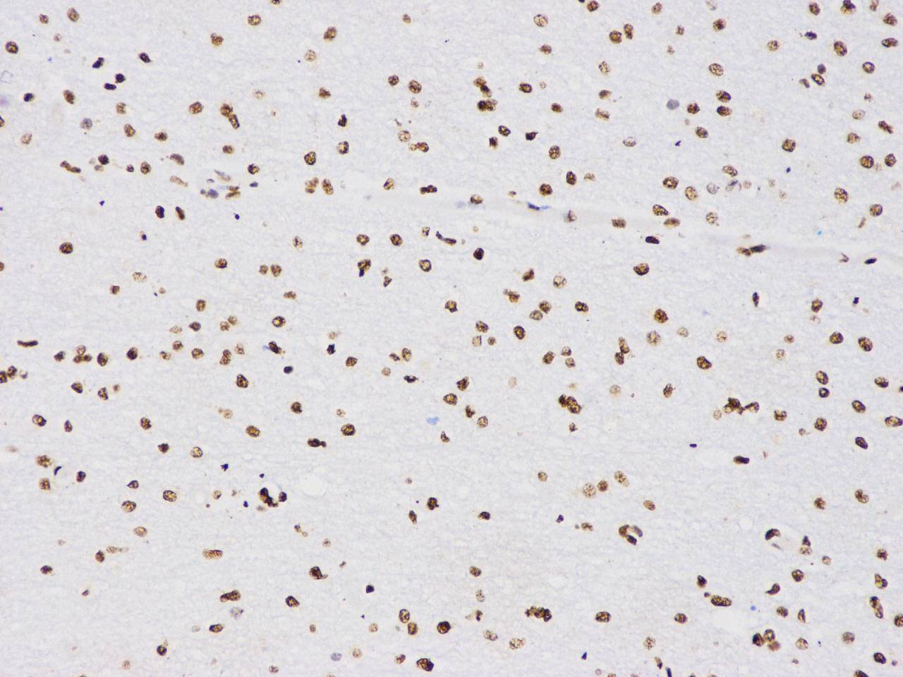
Fig3:Immunohistochemical analysis of paraffin-embedded human brain tissue using anti-ESRRB rabbit polyclonal antibody.Application
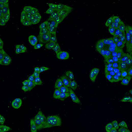
Fig4: ICC staining ESRRB in Hela cells (green). Cells were fixed in paraformaldehyde, permeabilised with 0.25% Triton X100/PBS and counterstained with DAPI in order to highlight the nucleus (blue).| Product Name | ESRRB |
|---|---|
| Antibody Type | Primary Antibodies |
| Antigen Alias | Err 2 antibody ERR b antibody ERR B2 antibody ERR beta 2 antibody ERR beta antibody ERR beta-2 antibody ERR-beta antibody Err2 antibody ERR2_HUMAN antibody ERRB 2 antibody ERRb antibody ERRB2 antibody ERRbeta 2 antibody ERRbeta antibody ESR L2 antibody ESRL 2 antibody ESRL2 antibody Esrrb antibody Estrogen receptor like 2 antibody Estrogen receptor related 2 antibody Estrogen receptor-like 2 antibody Estrogen-related receptor beta antibody Estrrb antibody Nr3b2 antibody Nuclear receptor ERRB2 antibody Nuclear receptor subfamily 3 group B member 2 antibody Orphan nuclear receptor antibody Steroid hormone receptor ERR 2 antibody Steroid hormone receptor ERR2 antibody |
| Product description | Steroid hormone receptors are found on the plasma membrane, in the cytosol and also in the nucleus of target cells. They are generally intracellular receptors and initiate signal transduction for steroid hormones which lead to changes in gene expression over a time period of hours to days. Defects in ESRRB are the cause of deafness autosomal recessive type 35 (DFNB35). DFNB35 is a form of sensorineural hearing loss. Sensorineural deafness results from damage to the neural receptors of the inner ear, the nerve pathways to the brain, or the area of the brain that receives sound information. |
| Immunogen | peptide |
| Clonality | Polyclonal |
|---|---|
| Isotype | IgG |
| Host Species | Rabbit |
| Tested Applications | WBICCIHC |
| WB:1:500-1:1,000 IHC:1:200 ICC:1:100-1:200 |
|
| Species Reactivity | HumanMouseRat |
| Concentration | 1mg/ml |
| Alternative Names | Err 2 antibody ERR b antibody ERR B2 antibody ERR beta 2 antibody ERR beta antibody ERR beta-2 antibody ERR-beta antibody Err2 antibody ERR2_HUMAN antibody ERRB 2 antibody ERRb antibody ERRB2 antibody ERRbeta 2 antibody ERRbeta antibody ESR L2 antibody ESRL 2 antibody ESRL2 antibody Esrrb antibody Estrogen receptor like 2 antibody Estrogen receptor related 2 antibody Estrogen receptor-like 2 antibody Estrogen-related receptor beta antibody Estrrb antibody Nr3b2 antibody Nuclear receptor ERRB2 antibody Nuclear receptor subfamily 3 group B member 2 antibody Orphan nuclear receptor antibody Steroid hormone receptor ERR 2 antibody Steroid hormone receptor ERR2 antibody |
|---|---|
| Molecular Weight(MW) | ~56 kDa |
| Cellular Localization | Nucleus |
| SwissProt ID | O95718 |
|---|
Application
Fig1: Western blot analysis on A549 (1) and F9 (2) cell lysates using anti-ESRRB rabbit polyclonal antibodies.
Application
Fig2:Immunohistochemical analysis of paraffin-embedded rat brain tissue using anti- ESRRB rabbit polyclonal antibody.
Application
Fig3:Immunohistochemical analysis of paraffin-embedded human brain tissue using anti-ESRRB rabbit polyclonal antibody.
Application
Fig4: ICC staining ESRRB in Hela cells (green). Cells were fixed in paraformaldehyde, permeabilised with 0.25% Triton X100/PBS and counterstained with DAPI in order to highlight the nucleus (blue).| Positive Control | F9, A549, human brain, rat brain, Hela |
|---|---|
| Application Notes | WB:1:500-1:1,000 IHC:1:200 ICC:1:100-1:200 |
| Form | Liquid |
|---|---|
| Storage Instructions | Store at +4℃ after thawing. Aliquot store at -20℃ or -80℃. Avoid repeated freeze / thaw cycles. |
| Storage Buffer | 1*TBS (pH7.4), 1%BSA, 40%Glycerol. Preservative: 0.05% Sodium Azide. |


