Application
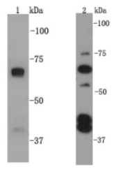
Fig1: Western blot analysis on Hela (1) and F9 (2) cell lysates using anti-SQSTM1 rabbit polyclonal antibodies.Application
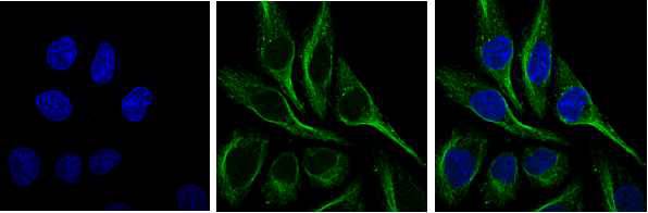
Fig2: ICC staining SQSTM1 in HepG2 cells (green). Cells were fixed in paraformaldehyde, permeabilised with 0.25% Triton X100/PBS and counterstained with DAPI in order to highlight the nucleus (blue).Application
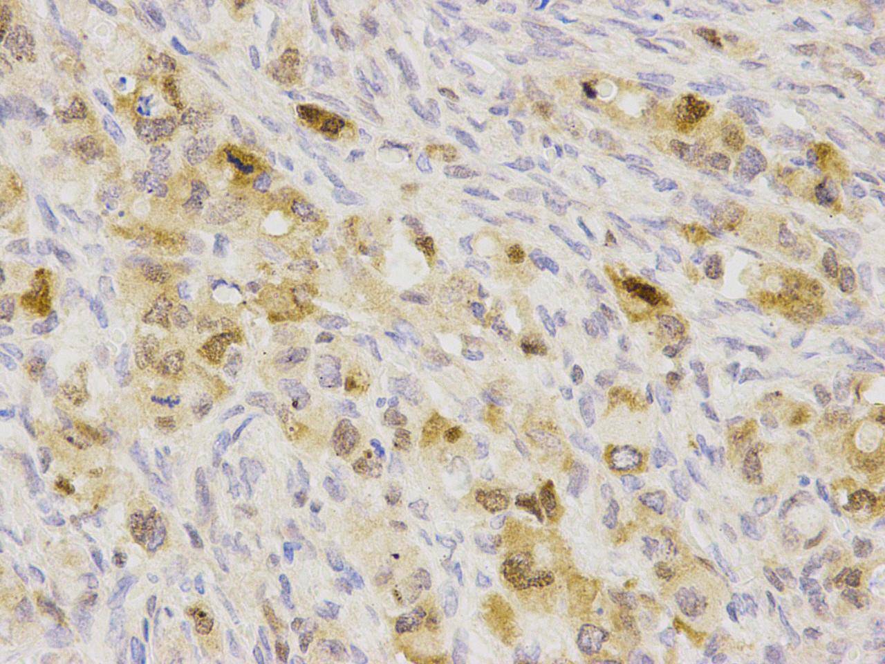
Fig3: Immunohistochemical analysis of paraffin-embedded human ovary carcinoma tissue using anti- SQSTM1 rabbit polyclonal antibody.| Product Name | SQSTM1 |
|---|---|
| Antibody Type | Primary Antibodies |
| Antigen Alias | A170 antibody DMRV antibody EBI 3 associated protein of 60 kDa antibody EBI 3 associated protein p60 antibody EBI3 associated protein of 60 kDa antibody EBI3 associated protein p60 antibody EBI3-associated protein of 60 kDa antibody EBIAP antibody FTDALS3 antibody MGC127197 antibody ORCA antibody OSF-6 antibody Osi antibody OSIL antibody Oxidative stress induced like antibody p60 antibody p62 antibody p62B antibody Paget disease of bone 3 antibody PDB 3 antibody PDB3 antibody Phosphotyrosine independent ligand for the Lck SH2 domain of 62 kDa antibody Phosphotyrosine independent ligand for the Lck SH2 domain p62 antibody Phosphotyrosine-independent ligand for the Lck SH2 domain of 62 kDa antibody PKC-zeta-interacting protein antibody Protein kinase C-zeta-interacting protein antibody Sequestosome 1 antibody Sequestosome-1 antibody SQSTM 1 antibody SQSTM_HUMAN antibody Sqstm1 antibody STAP antibody STONE14 antibody Ubiquitin binding protein p62 antibody Ubiquitin-binding protein p62 antibody ZIP 3 antibody ZIP antibody ZIP3 antibody |
| Product description | Sequestosome 1 (SQSTM1, p62) is a ubiquitin binding protein involved in cell signaling, oxidative stress and autophagy. It was first identified as a protein that binds to the SH2 domain of p56Lck and independently found to interact with PKCζ. SQSTM1 was subsequently found to interact with ubiquitin, providing a scaffold for several signaling proteins and triggering degradation of proteins through the proteasome or lysosome. Interaction between SQSTM1 and TRAF6 leads to the K63-linked polyubiquitination of TRAF6 and subsequent activation of the NF-κB pathway. It may play a role in titin/TTN downstream signaling in muscle cells and regulate signaling cascades through ubiquitination. P62 may be involved in cell differentiation, apoptosis, immune response and regulation of K+ channels. |
| Immunogen | peptide |
| Clonality | Polyclonal |
|---|---|
| Isotype | IgG |
| Host Species | Rabbit |
| Tested Applications | WBICCIHC |
| WB:1:500-1:1,000 ICC:1:200 IHC :1:200: |
|
| Species Reactivity | HumanMouseRat |
| Concentration | 1mg/ml |
| Alternative Names | A170 antibody DMRV antibody EBI 3 associated protein of 60 kDa antibody EBI 3 associated protein p60 antibody EBI3 associated protein of 60 kDa antibody EBI3 associated protein p60 antibody EBI3-associated protein of 60 kDa antibody EBIAP antibody FTDALS3 antibody MGC127197 antibody ORCA antibody OSF-6 antibody Osi antibody OSIL antibody Oxidative stress induced like antibody p60 antibody p62 antibody p62B antibody Paget disease of bone 3 antibody PDB 3 antibody PDB3 antibody Phosphotyrosine independent ligand for the Lck SH2 domain of 62 kDa antibody Phosphotyrosine independent ligand for the Lck SH2 domain p62 antibody Phosphotyrosine-independent ligand for the Lck SH2 domain of 62 kDa antibody PKC-zeta-interacting protein antibody Protein kinase C-zeta-interacting protein antibody Sequestosome 1 antibody Sequestosome-1 antibody SQSTM 1 antibody SQSTM_HUMAN antibody Sqstm1 antibody STAP antibody STONE14 antibody Ubiquitin binding protein p62 antibody Ubiquitin-binding protein p62 antibody ZIP 3 antibody ZIP antibody ZIP3 antibody |
|---|---|
| Molecular Weight(MW) | 62kDa |
| Cellular Localization | Cytoplasm |
| SwissProt ID | Q13501 |
|---|
Application
Fig1: Western blot analysis on Hela (1) and F9 (2) cell lysates using anti-SQSTM1 rabbit polyclonal antibodies.
Application
Fig2: ICC staining SQSTM1 in HepG2 cells (green). Cells were fixed in paraformaldehyde, permeabilised with 0.25% Triton X100/PBS and counterstained with DAPI in order to highlight the nucleus (blue).
Application
Fig3: Immunohistochemical analysis of paraffin-embedded human ovary carcinoma tissue using anti- SQSTM1 rabbit polyclonal antibody.| Positive Control | Hela, F9, HepG2, human ovary carcinoma tissue |
|---|---|
| Application Notes | WB:1:500-1:1,000 ICC:1:200 IHC :1:200: |
| Form | Liquid |
|---|---|
| Storage Instructions | Store at +4℃ after thawing. Aliquot store at -20℃ or -80℃. Avoid repeated freeze / thaw cycles. |
| Storage Buffer | 1*TBS (pH7.4), 1%BSA, 40%Glycerol. Preservative: 0.05% Sodium Azide. |


