Application
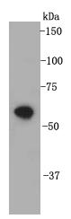
Fig1: Western blot analysis on cell lysates using anti- CD332 rabbit polyclonal antibodies.Application
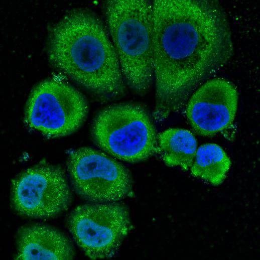
Fig2: ICC staining CD332 in SK-BR-3 cells (green). Cells were fixed in paraformaldehyde, permeabilised with 0.25% Triton X100/PBS and counterstained with DAPI in order to highlight the nucleus (blue).| Product Name | FGFR2/CD332 |
|---|---|
| Antibody Type | Primary Antibodies |
| Antigen Alias | bacteria-expressed kinase antibody BBDS antibody BEK antibody BEK fibroblast growth factor receptor antibody BFR1 antibody CD332 antibody CD332 antigen antibody CEK3 antibody CFD1 antibody Craniofacial dysostosis 1 antibody ECT1 antibody FGF receptor antibody FGFR 2 antibody FGFR-2 antibody Fgfr2 antibody FGFR2_HUMAN antibody Fibroblast growth factor receptor 2 antibody Hydroxyaryl protein kinase antibody Jackson Weiss syndrome antibody JWS antibody K SAM antibody K-sam antibody Keratinocyte growth factor receptor 2 antibody Keratinocyte growth factor receptor antibody KGFR antibody KSAM antibody protein tyrosine kinase, receptor like 14 antibody soluble FGFR4 variant 4 antibody TK14 antibody TK25 antibody |
| Product description | Fibroblast growth factor receptor 2 (FGFR2) also known as CD332 has two naturally occurring isoforms FGFR2IIIb and FGFR2IIIc, created by splicing of the third immunoglobulin-like domain. FGFR2IIIb is predominantly found in ectoderm derived tissues and endothelial organ lining, i.e. skin and internal organs. FGFR2 has important roles in embryonic development and tissue repair, especially bone and blood vessels. Like the other members of the Fibroblast growth factor receptor family, these receptors signal by binding to their ligand and dimerisation (pairing of receptors), which causes the tyrosine kinase domains to initiate a cascade of intracellular signals. As mentioned, FGFR2 mutations are associated with craniosynostosis syndromes, which are skull malformations caused by premature fusion of cranial sutures and other disease features according to the mutation itself. |
| Immunogen | peptide |
| Clonality | Polyclonal |
|---|---|
| Isotype | IgG |
| Host Species | Rabbit |
| Tested Applications | WBICCIHCFC |
| WB:1:2,000 ICC:1:200 |
|
| Species Reactivity | Human |
| Concentration | 1mg/ml |
| Alternative Names | bacteria-expressed kinase antibody
BBDS antibody
BEK antibody
BEK fibroblast growth factor receptor antibody
BFR1 antibody
CD332 antibody
CD332 antigen antibody
CEK3 antibody
CFD1 antibody
Craniofacial dysostosis 1 antibody
ECT1 antibody
FGF receptor antibody
FGFR 2 antibody
FGFR-2 antibody
Fgfr2 antibody
FGFR2_HUMAN antibody
Fibroblast growth factor receptor 2 antibody
Hydroxyaryl protein kinase antibody
Jackson Weiss syndrome antibody
JWS antibody
K SAM antibody
K-sam antibody
Keratinocyte growth factor receptor 2 antibody
Keratinocyte growth factor receptor antibody
KGFR antibody
KSAM antibody
protein tyrosine kinase receptor like 14 antibody soluble FGFR4 variant 4 antibody TK14 antibody TK25 antibody |
|---|---|
| Molecular Weight(MW) | ~100 kDa |
| Cellular Localization | Cell membrane, golgi apparatus, cytoplasmic vesicle |
| SwissProt ID | P21802 |
|---|
Application
Fig1: Western blot analysis on cell lysates using anti- CD332 rabbit polyclonal antibodies.
Application
Fig2: ICC staining CD332 in SK-BR-3 cells (green). Cells were fixed in paraformaldehyde, permeabilised with 0.25% Triton X100/PBS and counterstained with DAPI in order to highlight the nucleus (blue).| Positive Control | MCF-7, K562, Jurkat |
|---|---|
| Application Notes | WB:1:2,000 ICC:1:200 |
| Form | Liquid |
|---|---|
| Storage Instructions | Store at +4℃ after thawing. Aliquot store at -20℃ or -80℃. Avoid repeated freeze / thaw cycles. |
| Storage Buffer | 1*TBS (pH7.4), 1%BSA, 40%Glycerol. Preservative: 0.05% Sodium Azide. |


