Application
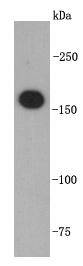
Fig1: Western blot analysis on Jurkat cell lysates using anti-TOP2A rabbit polyclonal antibodies.Application
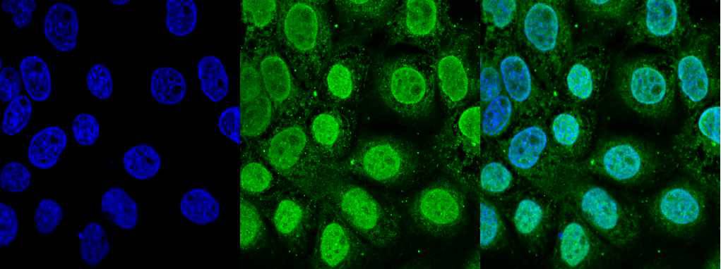
Fig2: ICC staining TOP2A in A431 cells (green). Cells were fixed in paraformaldehyde, permeabilised with 0.25% Triton X100/PBS and counterstained with DAPI in order to highlight the nucleus (blue).| Product Name | TOP2A |
|---|---|
| Antibody Type | Primary Antibodies |
| Antigen Alias | alpha isozyme antibody ATP hydrolyzing DNA topoisomerase II alfa antibody DNA gyrase antibody DNA topoisomerase (ATP hydrolyzing) antibody DNA topoisomerase 2 alpha antibody DNA topoisomerase 2-alpha antibody DNA topoisomerase II 170 kD antibody DNA topoisomerase II alpha isozyme antibody DNA topoisomerase II antibody DNA Topoisomerase2 antibody TOP 2A antibody TOP2 antibody TOP2A antibody TOP2A_HUMAN antibody Topoisomerase DNA II alpha 170kDa antibody TP2A antibody |
| Product description | DNA topoisomerases I and II are nuclear enzymes; type II consists of two highly homologous isoforms: topoisomerase IIα and IIβ. This nuclear enzyme is involved in processes such as chromosome condensation, chromatid separation, and the relief of torsional stress that occurs during DNA transcription and replication. It catalyzes the transient breaking and rejoining of two strands of duplex DNA which allows the strands to pass through one another, thus altering the topology of DNA. Topoisomerases are very susceptible to various stresses. Acidic pH or oxidative stress can convert topoisomerases to DNA-breaking nucleases, causing genomic instability and cell death. DNA-damaging topoisomerase targeting drugs (e.g., etoposide) also convert topoisomerases to nucleases, with the enzyme usually trapped as an intermediate that is covalently bound to the 5+ end of the cleaved DNA strand(s). |
| Immunogen | recombinant protein |
| Clonality | Polyclonal |
|---|---|
| Isotype | IgG |
| Host Species | Rabbit |
| Tested Applications | WBICC |
| WB:1:1,000-1:2,000 ICC:1:200 |
|
| Species Reactivity | HumanMouseRat |
| Concentration | 1mg/ml |
| Alternative Names | alpha isozyme antibody ATP hydrolyzing DNA topoisomerase II alfa antibody DNA gyrase antibody DNA topoisomerase (ATP hydrolyzing) antibody DNA topoisomerase 2 alpha antibody DNA topoisomerase 2-alpha antibody DNA topoisomerase II 170 kD antibody DNA topoisomerase II alpha isozyme antibody DNA topoisomerase II antibody DNA Topoisomerase2 antibody TOP 2A antibody TOP2 antibody TOP2A antibody TOP2A_HUMAN antibody Topoisomerase DNA II alpha 170kDa antibody TP2A antibody |
|---|---|
| Molecular Weight(MW) | 175 kDa |
| Cellular Localization | Nucleus |
| SwissProt ID | Q01320 |
|---|
Application
Fig1: Western blot analysis on Jurkat cell lysates using anti-TOP2A rabbit polyclonal antibodies.
Application
Fig2: ICC staining TOP2A in A431 cells (green). Cells were fixed in paraformaldehyde, permeabilised with 0.25% Triton X100/PBS and counterstained with DAPI in order to highlight the nucleus (blue).| Positive Control | Jurkat, A431 |
|---|---|
| Application Notes | WB:1:1,000-1:2,000 ICC:1:200 |
| Form | Liquid |
|---|---|
| Storage Instructions | Store at +4℃ after thawing. Aliquot store at -20℃ or -80℃. Avoid repeated freeze / thaw cycles. |
| Storage Buffer | 1*TBS (pH7.4), 1%BSA, 40%Glycerol. Preservative: 0.05% Sodium Azide. |


