Application
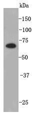
Fig1: Western blot analysis on hES cell lysates using anti-PRDM14 rabbit polyclonal antibodies.Application
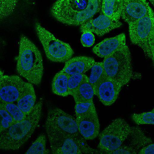
Fig2: ICC staining PRDM14 in MCF-7 cells (green). Cells were fixed in paraformaldehyde, permeabilised with 0.25% Triton X100/PBS and counterstained with DAPI in order to highlight the nucleus (blue).Application
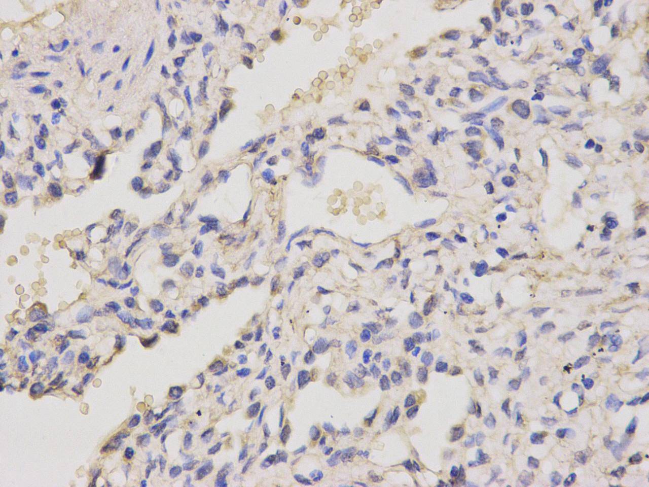
Fig3: Immunohistochemical analysis of paraffin-embedded human lung tissue using anti- PRDM14 rabbit polyclonal antibody.| Product Name | PRDM14 |
|---|---|
| Antibody Type | Primary Antibodies |
| Antigen Alias | MGC59730 antibody PFM 11 antibody PFM11 antibody PR domain containing 14 antibody PR domain zinc finger protein 14 antibody PR domain-containing protein 14 antibody PRD14_HUMAN antibody PRDM 14 antibody PRDM14 antibody |
| Product description | PR domain zinc finger protein 14 is a transcription factor that has both positive and negative roles on transcription. It is expressed in embryonic stem cells and tends to be overexpressed in breast cancer. PRDM14 is required for the maintenance of emryonic stem cell identity and the reacquisition of pluripotency in somatic cells. It may play an essential role in germ cell development at 2 levels: the reacquisition of potential pluripotency, including SOX2 up-regulation, and successful epigenetic reprogramming, characterized by EHMT1 repression. PRDM14 directly up-regulates the expression of pluripotency gene POU5F1 through its proximal enhancer. |
| Immunogen | peptide |
| Clonality | Polyclonal |
|---|---|
| Isotype | IgG |
| Host Species | Rabbit |
| Tested Applications | WBICCIHC |
| WB:1:500-1:1,000 ICC:1:100: IHC: 1:100: |
|
| Species Reactivity | HumanMouse |
| Concentration | 1mg/ml |
| Alternative Names | MGC59730 antibody PFM 11 antibody PFM11 antibody PR domain containing 14 antibody PR domain zinc finger protein 14 antibody PR domain-containing protein 14 antibody PRD14_HUMAN antibody PRDM 14 antibody PRDM14 antibody |
|---|---|
| Molecular Weight(MW) | ~64kDa |
| Cellular Localization | Nucleus |
| SwissProt ID | Q9GZV8 |
|---|
Application
Fig1: Western blot analysis on hES cell lysates using anti-PRDM14 rabbit polyclonal antibodies.
Application
Fig2: ICC staining PRDM14 in MCF-7 cells (green). Cells were fixed in paraformaldehyde, permeabilised with 0.25% Triton X100/PBS and counterstained with DAPI in order to highlight the nucleus (blue).
Application
Fig3: Immunohistochemical analysis of paraffin-embedded human lung tissue using anti- PRDM14 rabbit polyclonal antibody.| Positive Control | Human embryonic stem cells, human lung, MCF-7 |
|---|---|
| Application Notes | WB:1:500-1:1,000 ICC:1:100: IHC: 1:100: |
| Form | Liquid |
|---|---|
| Storage Instructions | Store at +4℃ after thawing. Aliquot store at -20℃ or -80℃. Avoid repeated freeze / thaw cycles. |
| Storage Buffer | 1*TBS (pH7.4), 1%BSA, 40%Glycerol. Preservative: 0.05% Sodium Azide. |


