Application
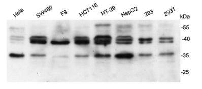
Fig1: Western blot analysis on different cell lysates using anti-Bag1 rabbit polyclonal antibodies.Application
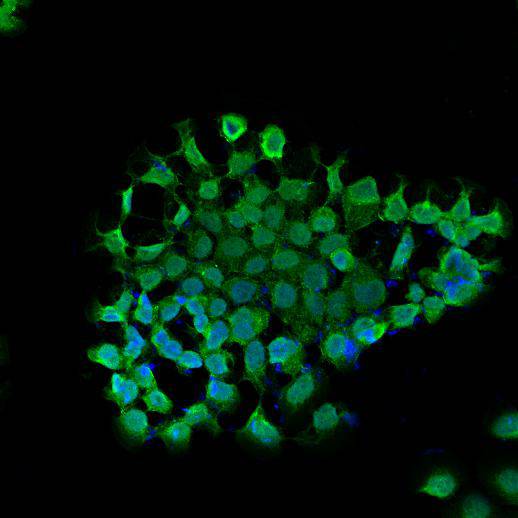
Fig2: ICC staining Bag1 in A431 cells (green). Cells were fixed in paraformaldehyde, permeabilised with 0.25% Triton X100/PBS and counterstained with DAPI in order to highlight the nucleus (blue).Application
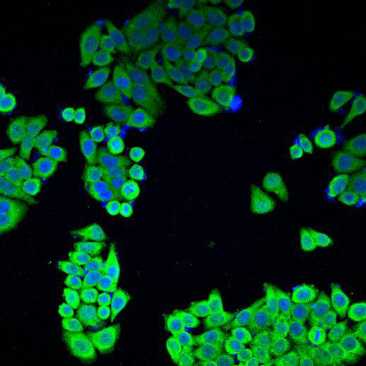
Fig3: ICC staining Bag1 in HepG2 cells (green). Cells were fixed in paraformaldehyde, permeabilised with 0.25% Triton X100/PBS and counterstained with DAPI in order to highlight the nucleus (blue).| Product Name | Bag1 |
|---|---|
| Antibody Type | Primary Antibodies |
| Antigen Alias | Bag 1 antibody BAG family molecular chaperone regulator 1 antibody BAG-1 antibody BAG1 antibody BAG1_HUMAN antibody BCL 2 Associated Athanogene 1 antibody BCL 2 associated athanogene antibody BCL 2 binding athanogene 1 antibody Bcl-2-associated athanogene 1 antibody BCL2 associated athanogene 1 antibody BCL2 associated athanogene antibody Glucocorticoid receptor-associated protein RAP46 antibody HAP1 antibody Haptoglobin antibody RAP 46 antibody RAP46 antibody |
| Product description | The oncogene BCL2 is a membrane protein that blocks a step in a pathway leading to apoptosis or programmed cell death. The protein encoded by this gene binds to BCL2 and is referred to as BCL2-associated athanogene. It enhances the anti-apoptotic effects of BCL2 and represents a link between growth factor receptors and anti-apoptotic mechanisms. At least three protein isoforms are encoded by this mRNA through the use of alternative translation initiation sites, including a non-AUG site. BAG gene has been implicated in age related neurodegenerative diseases as Alzheimer's. It has been demonstrated that BAG1 and BAG 3 regulate the proteasomal and lysosomal protein elimination pathways, respectively. |
| Immunogen | Recombinant protein within human Bag1 full sequence. |
| Clonality | Polyclonal |
|---|---|
| Isotype | IgG |
| Host Species | Rabbit |
| Tested Applications | WBICC |
| WB:1:1000 ICC:1:200 |
|
| Species Reactivity | HumanMouseRat |
| Concentration | 1mg/ml |
| Alternative Names | Bag 1 antibody BAG family molecular chaperone regulator 1 antibody BAG-1 antibody BAG1 antibody BAG1_HUMAN antibody BCL 2 Associated Athanogene 1 antibody BCL 2 associated athanogene antibody BCL 2 binding athanogene 1 antibody Bcl-2-associated athanogene 1 antibody BCL2 associated athanogene 1 antibody BCL2 associated athanogene antibody Glucocorticoid receptor-associated protein RAP46 antibody HAP1 antibody Haptoglobin antibody RAP 46 antibody RAP46 antibody |
|---|---|
| Molecular Weight(MW) | 35, 38 kDa |
| Cellular Localization | Cytoplasm. Nucleus |
| SwissProt ID | Q99933 |
|---|
Application
Fig1: Western blot analysis on different cell lysates using anti-Bag1 rabbit polyclonal antibodies.
Application
Fig2: ICC staining Bag1 in A431 cells (green). Cells were fixed in paraformaldehyde, permeabilised with 0.25% Triton X100/PBS and counterstained with DAPI in order to highlight the nucleus (blue).
Application
Fig3: ICC staining Bag1 in HepG2 cells (green). Cells were fixed in paraformaldehyde, permeabilised with 0.25% Triton X100/PBS and counterstained with DAPI in order to highlight the nucleus (blue).| Positive Control | Hela, SW480, F9, HCT116, HT-29, HepG2, 293, 293T, A431 |
|---|---|
| Application Notes | WB:1:1000 ICC:1:200 |
| Form | Liquid |
|---|---|
| Storage Instructions | Store at +4℃ after thawing. Aliquot store at -20℃ or -80℃. Avoid repeated freeze / thaw cycles. |
| Storage Buffer | 1*TBS (pH7.4), 1%BSA, 40%Glycerol. Preservative: 0.05% Sodium Azide. |


