Application
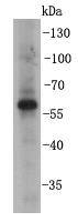
Fig1: Western blot analysis on SHG-44 cell lysates using anti- BMPR1B rabbit polyclonal antibodies.Application
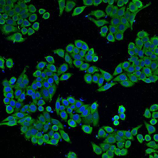
Fig2: ICC staining BMPR1B in HepG2 cells (green). Cells were fixed in paraformaldehyde, permeabilised with 0.25% Triton X100/PBS and counterstained with DAPI in order to highlight the nucleus (blue).Application
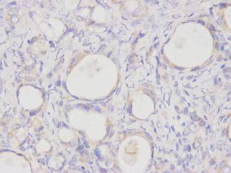
Fig3: Immunohistochemical analysis of paraffin-embedded human ovarian carcinoma tissue using anti- BMPR1B rabbit polyclonal antibody.| Product Name | BMPR1B |
|---|---|
| Antibody Type | Primary Antibodies |
| Antigen Alias | Activin receptor like kinase 6 antibody Acvrlk6 antibody ALK 6 antibody ALK6 antibody alk6tr antibody BMP type-1B receptor antibody BMPR IB antibody BMPR-1B antibody Bmpr1b antibody BMPRIB antibody BMR1B_HUMAN antibody Bone morphogenetic protein receptor type 1B antibody Bone morphogenetic protein receptor type IB antibody Bone morphogenetic protein receptor type-1B antibody BR 1b antibody BR1b antibody CDw 293 antibody CDw293 antibody CDw293 antigen antibody CFK 43a antibody CFK43a antibody Serine/threonine receptor kinase antibody zALK 6 antibody zALK6 antibody |
| Product description | BMPR1B is a member of the bone morphogenetic protein (BMP) receptor family of transmembrane serine/threonine kinases. The ligands of this receptor are BMPs, which are members of the TGF-beta superfamily. BMPs are involved in endochondral bone formation and embryogenesis. These proteins transduce their signals through the formation of heteromeric complexes of 2 different types of serine (threonine) kinase receptors: type I receptors of about 50-55 kD and type II receptors of about 70-80 kD. The BMPR1B receptor plays a role in the formation of middle and proximal phalanges. Mutations in this gene have been associated with primary pulmonary hypertension. In the chick embryo, it has been shown that BMPR1B is found in precartilaginous condensations. |
| Immunogen | peptide |
| Clonality | Polyclonal |
|---|---|
| Isotype | IgG |
| Host Species | Rabbit |
| Tested Applications | WBICCIHC |
| WB:1:1000 IHC:1:100 ICC:1:200 |
|
| Species Reactivity | HumanMouse |
| Concentration | 1mg/ml |
| Alternative Names | Activin receptor like kinase 6 antibody Acvrlk6 antibody ALK 6 antibody ALK6 antibody alk6tr antibody BMP type-1B receptor antibody BMPR IB antibody BMPR-1B antibody Bmpr1b antibody BMPRIB antibody BMR1B_HUMAN antibody Bone morphogenetic protein receptor type 1B antibody Bone morphogenetic protein receptor type IB antibody Bone morphogenetic protein receptor type-1B antibody BR 1b antibody BR1b antibody CDw 293 antibody CDw293 antibody CDw293 antigen antibody CFK 43a antibody CFK43a antibody Serine/threonine receptor kinase antibody zALK 6 antibody zALK6 antibody |
|---|---|
| Molecular Weight(MW) | 57kDa |
| Cellular Localization | Cell membrane |
| SwissProt ID | O00238 |
|---|
Application
Fig1: Western blot analysis on SHG-44 cell lysates using anti- BMPR1B rabbit polyclonal antibodies.
Application
Fig2: ICC staining BMPR1B in HepG2 cells (green). Cells were fixed in paraformaldehyde, permeabilised with 0.25% Triton X100/PBS and counterstained with DAPI in order to highlight the nucleus (blue).
Application
Fig3: Immunohistochemical analysis of paraffin-embedded human ovarian carcinoma tissue using anti- BMPR1B rabbit polyclonal antibody.| Positive Control | HepG2, SHG-44, human ovarian carcinoma tissue |
|---|---|
| Application Notes | WB:1:1000 IHC:1:100 ICC:1:200 |
| Form | Liquid |
|---|---|
| Storage Instructions | Store at +4℃ after thawing. Aliquot store at -20℃ or -80℃. Avoid repeated freeze / thaw cycles. |
| Storage Buffer | 1*TBS (pH7.4), 1%BSA, 40%Glycerol. Preservative: 0.05% Sodium Azide. |


