Application
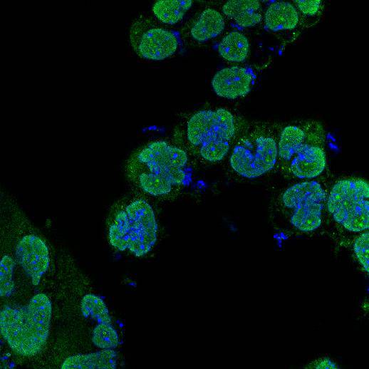
Fig1: ICC staining Geminin in F9 cells (green). Cells were fixed in paraformaldehyde, permeabilised with 0.25% Triton X100/PBS and counterstained with DAPI in order to highlight the nucleus (blue).Application
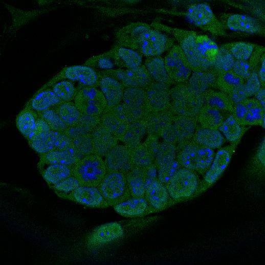
Fig2: ICC staining Geminin in D3 cells (green). Cells were fixed in paraformaldehyde, permeabilised with 0.25% Triton X100/PBS and counterstained with DAPI in order to highlight the nucleus (blue).Application
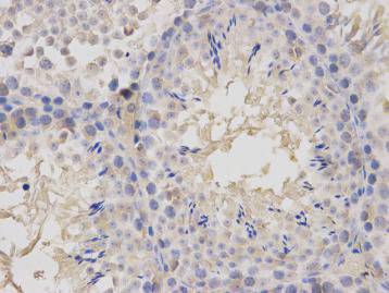
Fig3: Immunohistochemical analysis of paraffin-embedded mouse testis tissue using anti- Geminin rabbit polyclonal antibody.| Product Name | Geminin |
|---|---|
| Antibody Type | Primary Antibodies |
| Antigen Alias | DNA replication inhibitor antibody Gem antibody GEMI_HUMAN antibody Geminin antibody Geminin DNA replication inhibitor antibody GMNN antibody OTTHUMP00000039393 antibody RP3 369A17.3 antibody |
| Product description | Geminin is a nuclear protein that is present in most eukaryotics and highly conserved across species. Numerous functions have been elucidated for Geminin including roles in metazoan cell cycle, cellular proliferation, cell lineage commitment, and neural differentiation. Geminin is absent during G1 phase and accumulates through S, G2 phase and M phases of the cell cycle. Geminin levels drop at the metaphase / anaphase transition of mitosis when it is degraded by the Anaphase Promoting Complex (APC/C). During S phase, geminin is a positive regulator of DNA replication. In many cancer cell lines, inhibition of geminin by RNAi results in re-replication of portions of the genome, which leads to aneuploidy. In these cell lines, geminin knockdown leads to markedly slowed growth and apoptosis within several days. Recently, geminin has been found to be overexpressed in several malignancies and cancer cell lines. |
| Immunogen | recombinant protein |
| Clonality | Polyclonal |
|---|---|
| Isotype | IgG |
| Host Species | Rabbit |
| Tested Applications | ICCIHCFC |
| IHC:1:100-1:200 ICC:1:200 |
|
| Species Reactivity | Mouse |
| Concentration | 1mg/ml |
| Alternative Names | DNA replication inhibitor antibody Gem antibody GEMI_HUMAN antibody Geminin antibody Geminin DNA replication inhibitor antibody GMNN antibody OTTHUMP00000039393 antibody RP3 369A17.3 antibody |
|---|---|
| Molecular Weight(MW) | ~23 kDa |
| Cellular Localization | Cytoplasm, nucleus |
| SwissProt ID | O88513 |
|---|
Application
Fig1: ICC staining Geminin in F9 cells (green). Cells were fixed in paraformaldehyde, permeabilised with 0.25% Triton X100/PBS and counterstained with DAPI in order to highlight the nucleus (blue).
Application
Fig2: ICC staining Geminin in D3 cells (green). Cells were fixed in paraformaldehyde, permeabilised with 0.25% Triton X100/PBS and counterstained with DAPI in order to highlight the nucleus (blue).
Application
Fig3: Immunohistochemical analysis of paraffin-embedded mouse testis tissue using anti- Geminin rabbit polyclonal antibody.| Positive Control | Mouse testis, D3, F9 |
|---|---|
| Application Notes | IHC:1:100-1:200 ICC:1:200 |
| Form | Liquid |
|---|---|
| Storage Instructions | Store at +4℃ after thawing. Aliquot store at -20℃ or -80℃. Avoid repeated freeze / thaw cycles. |
| Storage Buffer | 1*TBS (pH7.4), 1%BSA, 40%Glycerol. Preservative: 0.05% Sodium Azide. |


