Application
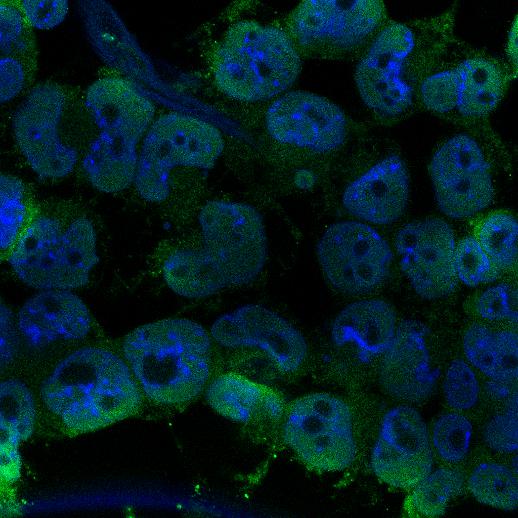
Fig1: ICC staining DPY30 in F9 cells (green). Cells were fixed in paraformaldehyde, permeabilised with 0.25% Triton X100/PBS and counterstained with DAPI in order to highlight the nucleus (blue).Application
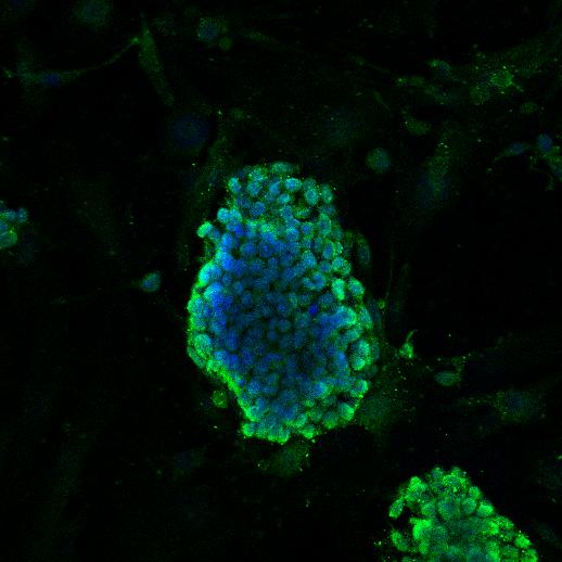
Fig2: ICC staining DPY30 in D3 cells (green). Cells were fixed in paraformaldehyde, permeabilised with 0.25% Triton X100/PBS and counterstained with DAPI in order to highlight the nucleus (blue).Application
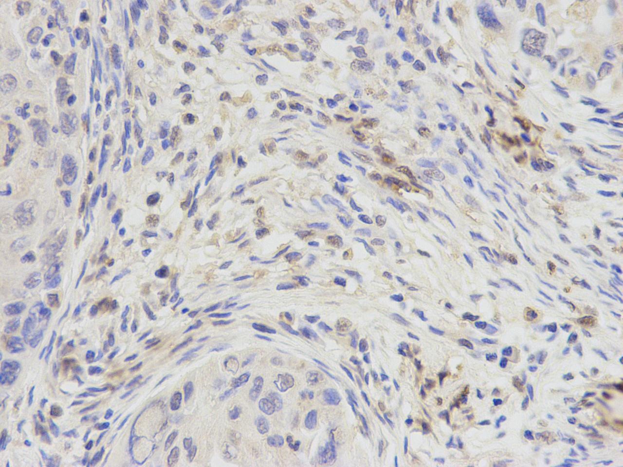
Fig3: Immunohistochemical analysis of paraffin-embedded human lung carcinoma tissue using anti- DPY30 rabbit polyclonal antibody.Application
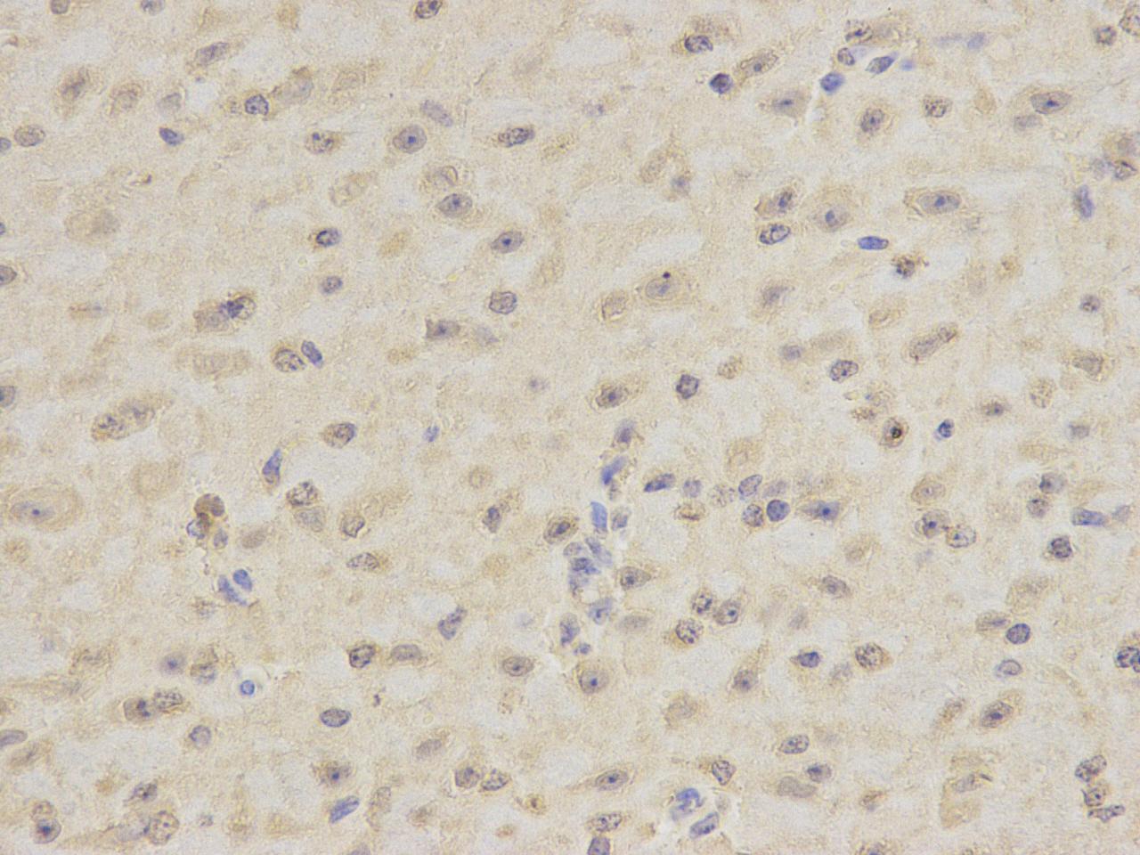
Fig4: Immunohistochemical analysis of paraffin-embedded mouse brain tissue using anti-DPY30 rabbit polyclonal antibody.| Product Name | DPY30 |
|---|---|
| Antibody Type | Primary Antibodies |
| Antigen Alias | Dpy-30-like protein antibody Dpy-30L antibody DPY30 antibody DPY30_HUMAN antibody HDPY-30 antibody Protein dpy-30 homolog antibody Saf19 antibody |
| Product description | As part of the MLL1/MLL complex, DPY30 is involved in the methylation of histone H3 at 'Lys-4', particularly trimethylation. Histone H3 'Lys-4' methylation represents a specific tag for epigenetic transcriptional activation. DPY30 may play some role in histone H3 acetylation. In a teratocarcinoma cell, it plays a crucial role in retinoic acid-induced differentiation along the neural lineage, regulating gene induction and H3 'Lys-4' methylation at key developmental loci. DPY30 may also play an indirect or direct role in endosomal transport. In the recent study, it showed that depletion of Dpy-30 does not affect ESC self-renewal, but significantly alters the differentiation potential of ESCs, particularly along the neural lineage. |
| Immunogen | recombinant protein |
| Clonality | Polyclonal |
|---|---|
| Isotype | IgG |
| Host Species | Rabbit |
| Tested Applications | ICCIHC |
| IHC:1:100-1:200 ICC:1:100-1:200 |
|
| Species Reactivity | HumanMouse |
| Concentration | 1mg/ml |
| Alternative Names | Dpy-30-like protein antibody Dpy-30L antibody DPY30 antibody DPY30_HUMAN antibody HDPY-30 antibody Protein dpy-30 homolog antibody Saf19 antibody |
|---|---|
| Cellular Localization | Nucleus. Golgi apparatus |
| SwissProt ID | Q9C005 |
|---|
Application
Fig1: ICC staining DPY30 in F9 cells (green). Cells were fixed in paraformaldehyde, permeabilised with 0.25% Triton X100/PBS and counterstained with DAPI in order to highlight the nucleus (blue).
Application
Fig2: ICC staining DPY30 in D3 cells (green). Cells were fixed in paraformaldehyde, permeabilised with 0.25% Triton X100/PBS and counterstained with DAPI in order to highlight the nucleus (blue).
Application
Fig3: Immunohistochemical analysis of paraffin-embedded human lung carcinoma tissue using anti- DPY30 rabbit polyclonal antibody.
Application
Fig4: Immunohistochemical analysis of paraffin-embedded mouse brain tissue using anti-DPY30 rabbit polyclonal antibody.| Positive Control | D3, F9, human lung carcinoma tissue, mouse brain |
|---|---|
| Application Notes | IHC:1:100-1:200 ICC:1:100-1:200 |
| Form | Liquid |
|---|---|
| Storage Instructions | Store at +4℃ after thawing. Aliquot store at -20℃ or -80℃. Avoid repeated freeze / thaw cycles. |
| Storage Buffer | 1*TBS (pH7.4), 1%BSA, 40%Glycerol. Preservative: 0.05% Sodium Azide. |


