Application
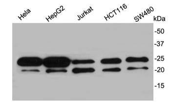
Fig1: Western blot analysis on different cell lysates using anti-Y14 rabbit polyclonal antibodies.Application
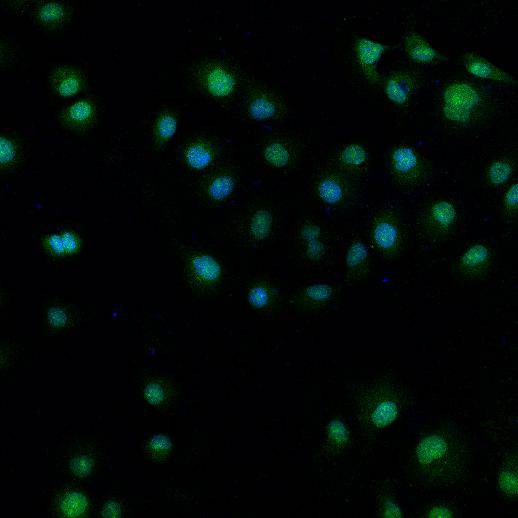
Fig2: ICC staining Y14 in HUVEC cells (green). Cells were fixed in paraformaldehyde, permeabilised with 0.25% Triton X100/PBS and counterstained with DAPI in order to highlight the nucleus (blue).Application
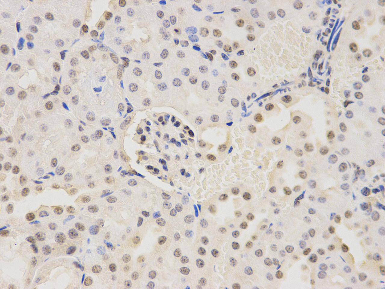
Fig3: Immunohistochemical analysis of paraffin-embedded mouse kidney tissue using anti- Y14 rabbit polyclonal antibody.Application
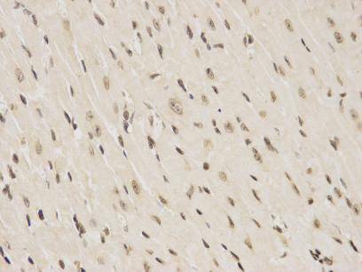
Fig4: Immunohistochemical analysis of paraffin-embedded mouse heart tissue using anti- Y14 rabbit polyclonal antibody.| Product Name | Y14 |
|---|---|
| Antibody Type | Primary Antibodies |
| Antigen Alias | Binder of OVCA1 1 antibody Binder of OVCA1-1 antibody BOV 1 antibody BOV 1A antibody BOV 1B antibody BOV 1C antibody BOV-1 antibody BOV1 antibody BOV1A antibody BOV1B antibody BOV1C antibody HSPC 114 antibody HSPC114 antibody MDS 014 antibody MDS014 antibody RBM 8 antibody RBM 8A antibody RBM 8B antibody RBM8 antibody rbm8a antibody RBM8A_HUMAN antibody RBM8B antibody Ribonucleoprotein RBM 8 antibody Ribonucleoprotein RBM 8A antibody Ribonucleoprotein RBM8 antibody Ribonucleoprotein RBM8A antibody RNA binding motif protein 8 antibody RNA binding motif protein 8A antibody RNA binding motif protein 8B antibody RNA binding protein 8A antibody RNA binding protein Y14 antibody RNA-binding motif protein 8A antibody RNA-binding protein 8A antibody RNA-binding protein Y14 antibody ZNRP antibody ZRNP 1 antibody ZRNP1 antibody |
| Product description | RNA-binding protein 8A is a protein that in humans is encoded by the RBM8A gene. This gene encodes a protein with a conserved RNA-binding motif. The protein is found predominantly in the nucleus, although it is also present in the cytoplasm. It is preferentially associated with mRNAs produced by splicing, including both nuclear mRNAs and newly exported cytoplasmic mRNAs. It is thought that the protein remains associated with spliced mRNAs as a tag to indicate where introns had been present, thus coupling pre- and post-mRNA splicing events. Previously, it was thought that two genes encode this protein, RBM8A and RBM8B; it is now thought that the RBM8B locus is a pseudogene. Two alternative start codons result in two forms of the protein, and this gene also uses multiple polyadenylation sites. |
| Immunogen | recombinant protein |
| Clonality | Polyclonal |
|---|---|
| Isotype | IgG |
| Host Species | Rabbit |
| Tested Applications | WBICCIHC |
| WB:1:2000-1:5000 IHC:1:200 ICC:1:200 |
|
| Species Reactivity | HumanMouseRat |
| Concentration | 1mg/ml |
| Alternative Names | Binder of OVCA1 1 antibody Binder of OVCA1-1 antibody BOV 1 antibody BOV 1A antibody BOV 1B antibody BOV 1C antibody BOV-1 antibody BOV1 antibody BOV1A antibody BOV1B antibody BOV1C antibody HSPC 114 antibody HSPC114 antibody MDS 014 antibody MDS014 antibody RBM 8 antibody RBM 8A antibody RBM 8B antibody RBM8 antibody rbm8a antibody RBM8A_HUMAN antibody RBM8B antibody Ribonucleoprotein RBM 8 antibody Ribonucleoprotein RBM 8A antibody Ribonucleoprotein RBM8 antibody Ribonucleoprotein RBM8A antibody RNA binding motif protein 8 antibody RNA binding motif protein 8A antibody RNA binding motif protein 8B antibody RNA binding protein 8A antibody RNA binding protein Y14 antibody RNA-binding motif protein 8A antibody RNA-binding protein 8A antibody RNA-binding protein Y14 antibody ZNRP antibody ZRNP 1 antibody ZRNP1 antibody |
|---|---|
| Molecular Weight(MW) | 20kDa |
| Cellular Localization | Nucleus |
| SwissProt ID | Q9Y5S9 |
|---|
Application
Fig1: Western blot analysis on different cell lysates using anti-Y14 rabbit polyclonal antibodies.
Application
Fig2: ICC staining Y14 in HUVEC cells (green). Cells were fixed in paraformaldehyde, permeabilised with 0.25% Triton X100/PBS and counterstained with DAPI in order to highlight the nucleus (blue).
Application
Fig3: Immunohistochemical analysis of paraffin-embedded mouse kidney tissue using anti- Y14 rabbit polyclonal antibody.
Application
Fig4: Immunohistochemical analysis of paraffin-embedded mouse heart tissue using anti- Y14 rabbit polyclonal antibody.| Positive Control | Hela, MCF-7, PANC-1, HepG2, HUVEC |
|---|---|
| Application Notes | WB:1:2000-1:5000 IHC:1:200 ICC:1:200 |
| Form | Liquid |
|---|---|
| Storage Instructions | Store at +4℃ after thawing. Aliquot store at -20℃ or -80℃. Avoid repeated freeze / thaw cycles. |
| Storage Buffer | 1*TBS (pH7.4), 1%BSA, 40%Glycerol. Preservative: 0.05% Sodium Azide. |


