Application
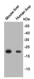
Fig1: Western blot analysis on different tissue lysates using anti- Glutathione peroxidase 1 rabbit polyclonal antibodies.Application
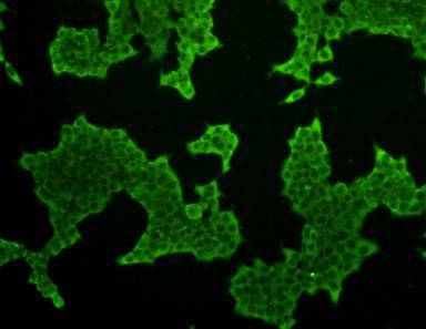
Fig2: ICC staining Glutathione peroxidase 1 in 293T cells (green). Cells were fixed in paraformaldehyde, permeabilised with 0.25% Triton X100/PBS.| Product Name | Glutathione peroxidase 1 |
|---|---|
| Antibody Type | Primary Antibodies |
| Antigen Alias | AL033363 antibody Cellular glutathione peroxidase antibody Glutathione peroxidase 1 antibody Glutathione peroxidase antibody GPx 1 antibody GPx-1 antibody GPX1 antibody GPX1_HUMAN antibody GPXD antibody GSHPx-1 antibody GSHPX1 antibody MGC14399 antibody MGC88245 antibody |
| Product description | Glutathione peroxidase 1 also known as GPx-1 is an enzyme that in humans is encoded by the GPX1 gene. Two alternatively spliced transcript variants encoding distinct isoforms have been found for this gene. This gene encodes a member of the glutathione peroxidase family, consisting of eight known glutathione peroxidases (Gpx1-8) in humans. Glutathione peroxidase functions in the detoxification of hydrogen peroxide, and is one of the most important antioxidant enzymes in humans. Glutathione peroxidase 1 is characterized in a polyalanine sequence polymorphism in the N-terminal region, which includes three alleles with five, six or seven alanine (Ala) repeats in this sequence. The allele with five Ala repeats is significantly associated with breast cancer risk. |
| Immunogen | peptide |
| Clonality | Polyclonal |
|---|---|
| Isotype | IgG |
| Host Species | Rabbit |
| Tested Applications | WBICCIHC |
| WB:1:500-1:1,000 ICC:1:100-1:200 |
|
| Species Reactivity | HumanMouse |
| Concentration | 1mg/ml |
| Alternative Names | AL033363 antibody Cellular glutathione peroxidase antibody Glutathione peroxidase 1 antibody Glutathione peroxidase antibody GPx 1 antibody GPx-1 antibody GPX1 antibody GPX1_HUMAN antibody GPXD antibody GSHPx-1 antibody GSHPX1 antibody MGC14399 antibody MGC88245 antibody |
|---|---|
| Molecular Weight(MW) | 22 kDa |
| Cellular Localization | Cytoplasm |
| SwissProt ID | P07203 |
|---|
Application
Fig1: Western blot analysis on different tissue lysates using anti- Glutathione peroxidase 1 rabbit polyclonal antibodies.
Application
Fig2: ICC staining Glutathione peroxidase 1 in 293T cells (green). Cells were fixed in paraformaldehyde, permeabilised with 0.25% Triton X100/PBS.| Positive Control | Human liver, mouse liver, 293T |
|---|---|
| Application Notes | WB:1:500-1:1,000 ICC:1:100-1:200 |
| Form | Liquid |
|---|---|
| Storage Instructions | Store at +4℃ after thawing. Aliquot store at -20℃ or -80℃. Avoid repeated freeze / thaw cycles. |
| Storage Buffer | 1*TBS (pH7.4), 0.5%BSA, 40%Glycerol. Preservative: 0.05% Sodium Azide. |


