Application
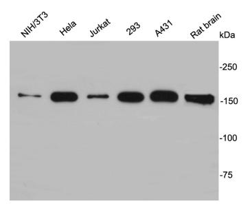
Fig1: Western blot analysis on different cell lysates using anti-EEA1 rabbit polyclonal antibodies.Application
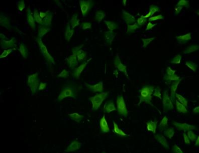
Fig2: ICC staining EEA1 in NIH/3T3 cells (green). Cells were fixed in paraformaldehyde, permeabilised with 0.25% Triton X100/PBS.| Product Name | EEA1 |
|---|---|
| Antibody Type | Primary Antibodies |
| Antigen Alias | Early endosome antigen 1 antibody Early endosome antigen 1, 162kD antibody Early endosome associated protein antibody EEA 1 antibody EEA1 antibody EEA1_HUMAN antibody Endosome associated protein p162 antibody Endosome-associated protein p162 antibody MST105 antibody MSTP105 antibody ZFYVE2 antibody Zinc finger FYVE domain containing protein 2 antibody Zinc finger FYVE domain-containing protein 2 antibody |
| Product description | EEA1 localizes exclusively to early endosomes and has an important role in endosomal trafficking. EEA1 binds directly to the phospholipid phosphatidylinositol 3-phosphate through its C-terminal FYVE domain and forms a homodimer through a coiled coil . EEA1 acts as a tethering molecule that couples vesicle docking with SNAREs such as N-ethylmaleimide sensitive fusion protein, bringing the endosomes physically closer and ultimately resulting in the fusion and delivery of endosomal cargo. Due to the proteins importance in vesicular trafficking, a number of intracellular bacteria prevent EEA1 recruitment to the vacuole. Mycobacterium tuberculosis is known to inhibit the recruitment of EEA1 to the phagosomal membrane through CamKII. |
| Immunogen | peptide |
| Clonality | Polyclonal |
|---|---|
| Isotype | IgG |
| Host Species | Rabbit |
| Tested Applications | WBICCIHC |
| WB:1:1000 ICC:1:100-1:200 |
|
| Species Reactivity | HumanMouseRat |
| Concentration | 1mg/ml |
| Alternative Names | Early endosome antigen 1 antibody
Early endosome antigen 1 162kD antibody Early endosome associated protein antibody EEA 1 antibody EEA1 antibody EEA1_HUMAN antibody Endosome associated protein p162 antibody Endosome-associated protein p162 antibody MST105 antibody MSTP105 antibody ZFYVE2 antibody Zinc finger FYVE domain containing protein 2 antibody Zinc finger FYVE domain-containing protein 2 antibody |
|---|---|
| Molecular Weight(MW) | 162kDa |
| Cellular Localization | Cytoplasm, early endosome membrane |
| SwissProt ID | Q15075 |
|---|
Application
Fig1: Western blot analysis on different cell lysates using anti-EEA1 rabbit polyclonal antibodies.
Application
Fig2: ICC staining EEA1 in NIH/3T3 cells (green). Cells were fixed in paraformaldehyde, permeabilised with 0.25% Triton X100/PBS.| Positive Control | Hela, Jurkat, NIH/3T3, 293, A431, rat brain |
|---|---|
| Application Notes | WB:1:1000 ICC:1:100-1:200 |
| Form | Liquid |
|---|---|
| Storage Instructions | Store at +4℃ after thawing. Aliquot store at -20℃ or -80℃. Avoid repeated freeze / thaw cycles. |
| Storage Buffer | 1*TBS (pH7.4), 1%BSA, 40%Glycerol. Preservative: 0.05% Sodium Azide. |


